
Sudden vision loss in one eye is a symptom that should not be ignored which also includes retinal vein occlusion as a very common cause in adults over fifty. To get an accurate retinal vein occlusion diagnosis is important because the earlier the blockage is identified the earlier a doctor can put together a treatment which in turn preserves what vision is left.
This article walks through everything from how retinal vein occlusion works to how doctors come up with a retinal vein occlusion diagnosis to what usually happens after that point when treatment planning begins. Just keep in mind, however, that while it’s good to understand as much about your condition as possible, nothing beats having an actual eye examination done by a professional.
What Is Retinal Vein Occlusion?
Retinal vein occlusion is a problem that occurs when one of the veins which drains blood out of the retina is blocked usually by a blood clot. The retina is the tissue at the back of the eye which is responsible for light sensitivity, and when it’s blood flow is reduced pressure increases in the smaller vessels which in turn causes fluid to spill into the retinal tissue and oxygen transport is impaired. The blockage is also most often associated with atherosclerosis which is the hardening and narrowing of the arteries that in turn press against a neighboring vein.
There are two main patterns this blockage can take, and understanding CRVO vs BRVO is central to understanding the condition as a whole.
CRVO vs BRVO: Two Different Patterns of Blockage
When it comes to CRVO vs BRVO what they are discussing is which part of the retinal venous system the blockage is at. In the case of Central retinal vein occlusion or CRVO the blockage is in the main vein which drains the entire retina so the effect is more widespread. With Branch retinal vein occlusion or BRVO the blockage is in a smaller branch vein which means the damage is usually seen in the section of the retina that that branch supplies.
| Feature | CRVO | BRVO |
| Location of blockage | Main central retinal vein | A smaller branch vein, often where an artery crosses over it |
| Typical extent of vision loss | Can affect vision across the whole eye | Often limited to part of the visual field |
| Relative frequency | Less common than BRVO | The more common of the two patterns |
| General outlook | More variable, sometimes more serious | Often somewhat better, especially with early treatment |
| Shared risk factors | High blood pressure, diabetes, atherosclerosis, glaucoma, clotting disorders | Same core risk factors as CRVO |
Both sets of patterns have the same base causes but the CRVO vs BRVO issue plays out different in how they are presented to doctors in terms of symptoms, diagnosis via imaging, and what results they should expect in terms of vision recovery.
What Causes Retinal Vein Occlusion?
Main risk factors for retinal vein occlusion are the same as that of in large for heart and vascular health. High blood pressure, diabetes, high cholesterol, and atherosclerosis are key in the development of the disease which in turn leaves the retinal veins more prone to blockage. Also included in this are other ophthalmic issues like glaucoma and past episodes of macular edema. Age is a factor which as we get older we see the progressive nature of these vascular changes which is why we see this in older people although it may present at a younger age if there is a hereditary clotting issue.
Recognizing the Symptoms
The main symptom of retinal vein occlusion is sudden painless blurring or loss of vision in one eye. In some people we note only a partial change which is for example blurriness in part of the visual field which is more common in BRVO, while in others we see a great deal more loss of vision as is reported in CRVO. As these symptoms may also present with other serious eye conditions we recommend a prompt evaluation instead of a wait and see approach.
How Is Retinal Vein Occlusion Diagnosed?
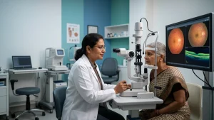
A proper retinal vein occlusion diagnosis relies on a combination of examination and imaging rather than a single test. An eye doctor typically starts with a dilated eye exam to directly view the retina and look for the hemorrhages, swelling, and dilated, twisted veins characteristic of a blockage.
| Diagnostic Step | What It Shows |
| Dilated eye exam | Direct view of retinal hemorrhages, swelling, and vein changes |
| Visual acuity test | Measures how much clarity of vision has been affected |
| Intraocular pressure check | Screens for related pressure changes, including secondary glaucoma |
| Fluorescein angiography | Uses a dye and specialized imaging to map blood flow and blocked areas |
| Optical coherence tomography, or OCT | Produces detailed cross sectional images of the retina to assess swelling |
| Blood tests | Checks for diabetes, cholesterol levels, and clotting disorders, especially in younger patients |
Reaching a confident retinal vein occlusion diagnosis often means monitoring the eye over several months as well, since certain complications, including glaucoma, can develop later rather than immediately after the initial blockage.
Planning Retina Blockage Treatment
Once the retinal vein occlusion diagnosis is confirmed, it is important to address issues like retinal swelling, and treating the health issues which caused the blockage in the first place.
Anti-VEGF injections are the primary modalities we use in retina blockage treatment at present. These drugs are put in the eye itself and what they do is they inhibit a protein called vascular endothelial growth factor which the retina overproduces when it is deprived of oxygen. By reducing this protein which plays a role in causing swelling and leakage the anti-VEGF agents play a role in vision improvement which is what we are after. Also we see that for many patients the results from the initial treatment phase of repeated anti-VEGF injections over a few months’ time may be long lasting. In many cases vision improvements from the use of anti-VEGF injections is sustained for a very long time beyond the immediate treatment period.
Corticosteroid infusions and implants also present an option for retina blockage treatment which do not respond fully to anti-VEGF injections. As for laser treatment which is what we term photocoagulation we also see this used for BRVO to decrease swelling or to deal with abnormal blood vessel growth in the retina.
Along in the wake of these direct treatments which do see a great deal of play; we also see a large focus put on what brought about the blockage in the first place. We do see success in terms of control of blood pressure, blood sugar, and cholesterol which in turn does not reverse the present damage, what we do see is that it does reduce the risk of the blockage progressing or affecting the other eye.
What Is the Outlook After Treatment?
The good news is that there are ways to treat each of these patterns effectively if they’re discovered and addressed quickly enough, but outcomes do vary quite a bit based on whether you have one form of retinal vein occlusion CRVO vs BRVO. In general, people who get the BRVO tend to have a slightly better prognosis than those with the CRVO variety.
Complications such as neovascular glaucoma might occur in more severe cases of either condition requiring further attention. Still, it is necessary to stay on top of things by making sure to attend all scheduled visits and stick with any therapy such as anti-VEGF injection treatments aimed at keeping your retina clear is important for maximizing the potential to maintain meaningful sight going forward regardless of which form presents itself.
When to Seek Immediate Care
Sudden changes in vision should not be put off until your routine eye exam. If you are experiencing blurry vision all of a sudden, a shadow that is like a curtain going over part of what you see, or if there is a quick decline in one of your eyesight, see your eye specialist as soon as you can. Early visit will give us the best chance at a proper retinal vein occlusion diagnosis, which in turn will play a role in the degree of vision recovery.
Conclusion
Retinal vein occlusion is a condition which presents itself when a blood clot, often a result of atherosclerosis, blocks the normal outflow of blood from the retina. In terms of the differential between CRVO vs BRVO, this is important in to note as it accounts for the great variation in symptoms and outcomes between patients. Also a detailed retinal vein occlusion diagnosis is a must which includes dilated eye exams, imaging, at time blood work before we begin any retina blockage treatment.
Anti-VEGF injections, corticosteroid options, laser treatment, and careful management of your base health issues all come into play for each individual case. If you see sudden changes in your vision which is not normal for you, get in to see your eye doctor as soon as you can which in turn gives you the best chance at protecting your sight.
Frequently Asked Questions
1. What is the main difference between CRVO vs BRVO?
In CRVO we see the primary retinal vein affected and this tends to present with large area involvement in terms of vision loss, while in BRVO a smaller branch vein is affected which in turn usually produces a smaller area of vision issue. Also they have similar risk factors which are high blood pressure and diabetes.
2. How long does retina blockage treatment usually take?
There is not a set time frame, that is determined by what the eye responds to. At first we do anti-VEGF injections which we give monthly and then we reduce the frequency as swelling goes down, some patients may require long term or even life time treatment.
3. Can retinal vein occlusion be prevented?
At present there is no guaranteed way to do so but what one can do is to manage blood pressure, blood sugar, and cholesterol, also to have regular eye exams which in turn may lower the risk, especially for people that have known cardiac risk factors.
4. Does vision always return after treatment?
No, not always. Some vision loss from retinal vein occlusion is permanent which is more likely if treatment is delayed. Many do see great improvement with early diagnosis and consistent treatment in which results may vary by the case.
5. Is retinal vein occlusion diagnosis painful?
No. Also it’s a fact that retinal vein occlusion diagnosis is not painful. A dilated eye exam, visual acuity test, and imaging like OCT or fluorescein angiography are not painful although the dilation drops will cause temporary light sensitivity and blurred near vision for a few hours.