
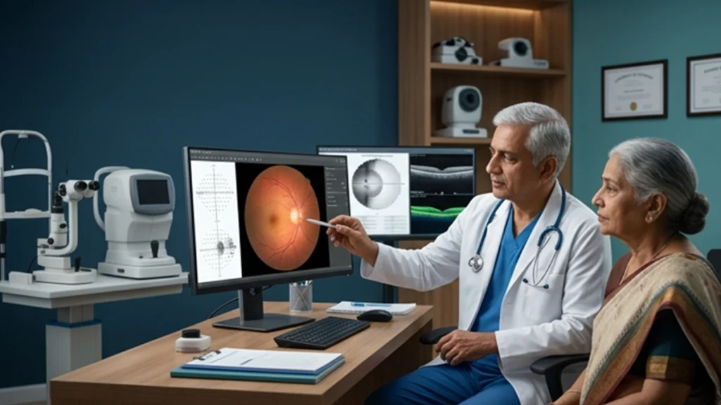
A report of a large cup disc ratio in the eye is at times very alarming. Many think of glaucoma first. But also a large cup does not in all cases point to disease. Some people are born with larger optic nerve cups. Also we see in some that certain changes which require close follow up. What we try to do is to put into perspective what is normal and what is not which in turn empowers patients to take better care of their eye health.
At ASG Eye Care, we see that regular eye exams can also catch early signs of nerve changes, in addition to that we’ll be able to know that it’s time for a more in depth test when required. We also note large cup disc ratio, that which we note as well as pressure in the eye, the eye’s structure and function.
What Does a Large Cup-to-Disc Ratio Mean?
The optic nerve which is responsible for carrying visual info from the eye to the brain. At the back of the eye is the optic disc which has a central defect we term the cup. The large cup to disc ratio is a term used to describe the size of this defect in relation to the whole of the optic disc.
A larger cup may be out of the norm and is a physical feature which in many cases is a result of a large optic disc. But also cup change is a sign of nerve fiber damage. Thus with respect to the cup in the optic nerve we must do a careful analysis and not jump to a diagnosis of glaucoma.
| Finding | What it may indicate |
| A large cup in a naturally large optic disc | Normal anatomical variation |
| A difference in cup size between the two eyes | A reason for closer assessment |
| Progressive cupping or thinning of the nerve rim | Possible glaucomatous damage |
| A large cup with other abnormal test results | Further glaucoma evaluation may be necessary |
The size of the cup alone cannot confirm or rule out glaucoma. The ophthalmologist considers the complete clinical picture.
Does Optic Nerve Cupping Always Mean Glaucoma?
No. Optic nerve cupping is a natural occurrence in some healthy eyes. Some people have a large cup which does not correspond to any loss of vision. Also we see progressive changes which may be indicative of glaucoma, a disease which affects the optic nerve and leads to permanent vision loss.
A person may be put into the category of glaucoma suspect when we see certain exam results or risk factors which raise red flags but at the same time do not give us a definitive diagnosis. This is not to say that glaucoma is a sure thing. What it does mean is that the eye in question requires a more in depth assessment and ongoing care.
A large cup disc ratio is of more concern when associated with thinning of the neuroretinal rim, changes in the retinal nerve fiber layer, high eye pressure or abnormal visual field results. Comparison of past notes will help the ophthalmologist to identify changes over time.
How Is the Condition Evaluated?
An ophthalmologist is able to study the optic nerve’s shape, cup size, rim tissue and appearance through an optic disc exam. Also we look at family history, age, intraocular pressure and other factors which play a role in glaucoma risk. For a full assessment many tests may be done. No single measurement gives all the answers which is why the results must be put together.
| Test | Purpose |
| Eye pressure measurement | Checks intraocular pressure |
| Dilated optic nerve assessment | Examines the optic disc and surrounding structures |
| Optical coherence tomography (OCT) | Measures nerve fibre layer and related structures |
| Visual field test | Checks for areas of reduced peripheral vision |
An optic disc examination may bring to notice features which require more in depth evaluation which in some cases may be in the absence of symptoms. Glaucoma does tend to develop very slowly which is why early assessment is of great value.
When Is Monitoring Important?
People who present with a large cup disc ratio may require regular checks up also if they have a family history of glaucoma, high eye pressure or what we term as suspicious OCT results. The interval between exams will vary according to the degree of concern and results of past tests.
For a glaucoma suspect we do so to see which way the optic nerve is going at the present time. Repetitive testing also brings to light very small changes which a single exam may not. A constant look over time may put into play a diagnosis of normal but at the same time the doctor has to look at all other issues.
Patients should go to all of their recommended appointments even when your vision appears normal. At early stages glaucoma may not produce noticeable symptoms and also by the time vision loss from damaged optic nerve occurs it is usually too late to reverse.
Conclusion
A large cup disc ratio is not in itself a sign of glaucoma. Some natural variation in optic nerve anatomy will present as such results, but progressive changes call for more in depth evaluation. Extensive optic disc examination, appropriate imaging and visual field test will determine the cause. It is very important to do a regular follow up for all which we suspect of having glaucoma. An ophthalmologist can clarify the results and will put into place the best plan for your care that will protect your long term vision.
Frequently Asked Questions
1. Is a large cup-to-disc ratio always abnormal?
No. Some people are born with larger optic nerve cups. We look at the size of the optic disc, nerve rim appearance, eye pressure and other tests to determine if the result is normal or suspicious.
2. Can a large cup-to-disc ratio indicate glaucoma?
Yes, that is one indicator of glaucoma but it isn’t definitive for the diagnosis. An ophthalmologist looks at other structural issues, visual field results and risk factors before coming to a decision.
3. Does a glaucoma suspect need treatment?
Not at all. Some patients do require monitoring as opposed to present treatment. What the ophthalmologist does is look at the chance of glumacoma, there is evidence of damage and what is seen at the follow up.
4. How often should the optic nerve be checked?
There isn’t a one size fits all schedule. The ophthalmologist recommends follow up based on individual risk, test results and if the optic nerve is changing over time.
References
For reliable patient information and clinical background, consult these authoritative sources:
- National Eye Institute (NEI) — National Eye Institute guidance on glaucoma detection and comprehensive eye examinations: https://www.nei.nih.gov/about/education-and-outreach/glaucoma-resources/national-eye-institute-statement-detection-glaucoma-and-adult-vision-screening
- American Academy of Ophthalmology (AAO) — Understanding glaucoma, its diagnosis and monitoring: https://www.aao.org/eye-health/diseases/what-is-glaucoma
- American Academy of Ophthalmology EyeWiki — Examination of the optic nerve: https://eyewiki.aao.org/Examination_of_the_optic_nerve_at_the_slit-lamp_biomicroscope_with_a_handheld_lens