Changes in the eye which are at the back may point out that there is a problem with the brain. Papilledema is a term we use for swelling of the optic disc which in turn is a result of increased pressure within the skull. Papilledema evaluation is a tool which doctors use to determine the cause of the issue and to assess the risk to vision. Early assessment is key because in cases which go untreated it may damage the optic nerve.
Not all cases of optic disc swelling are due to papilledema. That said we as health care providers have to look into the cause of raised intracranial pressure at the first sign of it even if the patient reports to be doing relatively well.
What Causes Papilledema?
The optic nerve at which the optic nerve connects to the retina is what we see with the optic disc. Also when brain pressure goes up it can affect this nerve and cause swelling. Papilledema evaluation is done to differentiate this from other eye or optic nerve issues.
Possible causes of this are a brain mass, inflammation, blocked cerebrospinal fluid drainage, or idiopathic intracranial hypertension. In the last case we see raised pressure with no identifiable cause. Raised intracranial pressure requires in depth investigation as the base cause may require urgent attention.
How Is Papilledema Evaluated?
1. Comprehensive Eye Exam
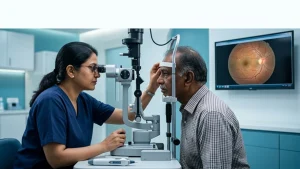
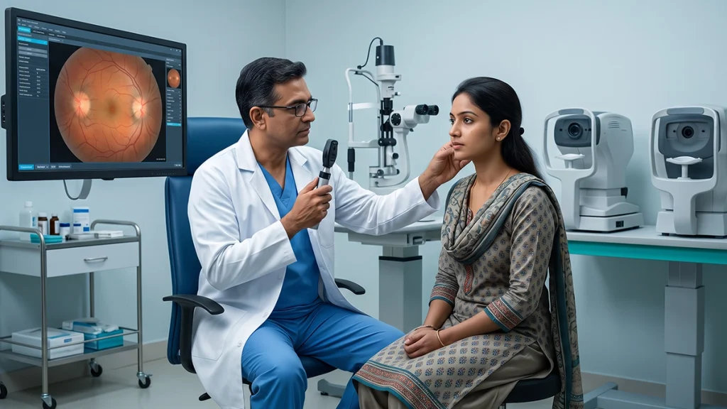
Doctors first go over medical history, medications, headaches, and changes in vision. In the case of papilledema evaluation we have eye specialists which dilate the pupil to look at the optic nerve. Also they may test visual acuity, check pupil response, and study peripheral vision.
During an exam we may note optic disc swelling, blurring of disc margins, or small hemorrhages. These are used by doctors to determine what further tests are needed.
2. Recognising Symptoms
Some at first present with headaches, nausea, or short lived episodes of dim vision. Also report of double vision and trouble seeing things at the periphery of vision. While these visual symptoms are a tell tale sign for some it is also true that some patients do not have much in the way of early warnings.
A papilledema evaluation includes symptoms and physical exam results. Profound headaches that are associated with vomit attacks, new onset of double vision, or sudden vision loss require immediate medical care.
3. Imaging the Brain
Doctors order up MRIs to determine the cause of what is thought to be raised intracranial pressure. Also they may perform MR venography which looks at the veins that drain blood from the brain. In a crisis setting a CT scan may be used.
Imaging is used to identify masses, fluid flow issues, or blood clots. For papilledema evaluation in turn may require coordination between ophthalmologists, neurologists, and other specialists.
4. Additional Diagnostic Tests
Optical coherence tomography (OCT) which we use to produce detailed images of the optic nerve and retina also allows us to document swelling and to monitor changes over time. For visual field testing we look at areas of reduced peripheral vision.
If as the case may be we do a lumbar puncture which follows brain imaging. This procedure measures cerebrospinal fluid pressure and also allows for analysis of the fluid. It is used to investigate raised intracranial pressure but of course we must first determine that the procedure is safe.
Common Tests and Their Purpose
| Test | Why doctors use it |
| Dilated eye examination | Identifies optic disc swelling and assesses the optic nerve. |
| OCT scan | Measures retinal nerve fibre layers and tracks changes. |
| Visual field test | Detects vision loss that ordinary reading charts may miss. |
| MRI or CT scan | Investigates possible causes inside the skull. |
| Lumbar puncture | Measures fluid pressure and examines cerebrospinal fluid when appropriate. |
Why Prompt Assessment Matters
Untreated papilledema may cause damage to the optic nerve and at times lead to permanent loss of vision. What that swelling is in degree of what is causing it, and the current state of the patient’s vision which in turn will play an important role in which treatment to which the patient is put. An papilledema evaluation is for the doctors to determine what the urgency level of the treatment required is.
| Warning sign | Recommended response |
| Sudden vision loss | Seek emergency medical care immediately. |
| Severe or worsening headache with vomiting | Obtain urgent medical assessment. |
| New double vision or repeated visual disturbances | Arrange prompt medical assessment. |
| Newly detected optic nerve swelling | Follow the specialist’s urgent investigation plan. |
Conclusion
Papilledema is just a symptom. It may present a serious issue which requires urgent attention. In it’s early papilledema evaluation can be a tell-tale sign for which we must do in depth investigation to determine the cause, to preserve vision, and to set out the best plan of care. If you see new changes in your vision or severe headaches report to a health care provider at once.
Frequently Asked Questions
1. Can papilledema present with no clear symptoms?
It can in some cases there are very mild or no early visual symptoms. Often the issue is detected during an eye exam which reveals the swelling before the person reports any change in vision.
2. Does it that every swollen optic disc is papilledema?
Not at all. Other conditions may present with that symptom. Doctors use of exam results, imaging and other tests to determine the cause.
3. Is papilledema a medical emergency?
It does present a serious underlying issue. In the case of suspected papilledema prompt assessment is required especially when there is raised intracranial pressure which may in turn threaten vision or health.
4. What goes into a papilledema evaluation?
During a papilledema evaluation work up a doctor may perform a dilated eye exam, OCT, visual field study, brain imaging, and a lumigray or spinal tap when appropriate. Which studies are done is dependent on what we find in each patient.
Government and Official Medical References
- National Library of Medicine, NIH: Papilledema — StatPearls
https://www.ncbi.nlm.nih.gov/books/NBK538295/ - NHS: Eye Injuries
https://www.nhs.uk/conditions/eye-injuries/