
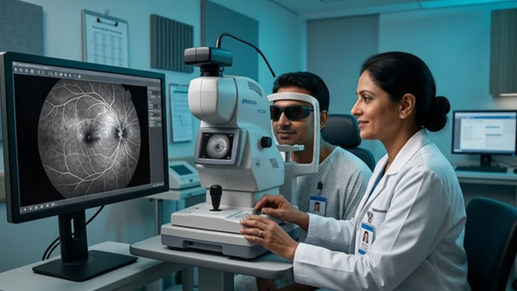
Changes at times present as issues in the small blood vessels at the back of the eye. These vessels may bleed, become blocked, or grow abnormally. A retinal angiography test which is used by ophthalmologists to get a detailed view of the changes and to see how blood moves through the retina.
Also known as the FFA Eye Test which is a type of fundus angiography, we use this investigation for very detailed images of the retinal blood vessels. Doctors may order it in which a routine eye exam doesn’t give us enough info about a retina issue.
What Is a Retinal Angiography Test?
A retinal angiography test is a diagnostic imaging which we use to look at circulation in the retina and choroid. For the test a health care provider will put fluorescein dye into a vein which is usually in the arm or hand. The dye gets into the blood stream and within seconds it goes to the eye.
A specialized retinal camera which in turn takes a series of images as the dye goes through the retinal blood vessels. From these images we are able to see areas that have reduced, blocked, abnormal, or leaking blood flow. The FFA eye test does not use X-rays. What it does use is certain light and filters to take a picture of the fluorescent dye as it moves around the eye.
| Aspect | What Happens |
| Preparation | Eye drops dilate the pupils for a clearer retinal view |
| Dye | Fluorescein dye enters a vein in the arm or hand |
| Imaging | A retinal camera photographs the dye moving through the eye |
| Purpose | The test identifies leakage, blockage, abnormal vessels and circulation changes |
| Results | The ophthalmologist reviews the images to guide diagnosis or treatment |
When Is an FFA Eye Test Recommended?
An ophthalmologist will put forward the idea of a retinal angiography test which presents itself as a solution when by what we see or what the symptom is telling us that there is a retinal vascular issue. The test in question will also be put forth as a choice when the doctor is trying to determine the site and degree of abnormal blood flow.
FFA eye test is a tool which helps in the evaluation of diabetic retinopathy, macular edema, retinal vein occlusion and certain types of age related macular degeneration. Also it plays a role in the assessment of inflammatory and vascular retinal diseases.
Diabetes can cause damage to the small retinal blood vessels which in turn may leak fluid or bleed. In advanced disease also it may happen that abnormal new vessels grow. The National Eye Institute reports that fluorescein angiography may be used when severe diabetic retinopathy or diabetic macular edema is suspected.
What Can a Retinal Angiography Test Detect?
The main advantage of a retinal angiography test is its ability to show circulation patterns that may not be obvious during a standard eye examination.
| Finding | What It May Indicate |
| Dye leakage | Damaged or abnormal blood vessels |
| Blocked circulation | Reduced retinal blood supply or vascular occlusion |
| Abnormal new vessels | Changes associated with conditions such as proliferative diabetic retinopathy |
| Areas of poor perfusion | Retinal tissue receiving inadequate blood flow |
| Macular leakage | Possible retinal swelling or macular edema |
Doctors which out of the fluorescein angiography results they look at how the dye fills and moves through the retinal circulation. We see in some cases leakage, delayed filling, poor flow, or atypical blood vessel patterns which in turn may help to identify the cause. A retinal angiography test also which in that it helps the ophthalmologist to see where the issue is at and also what the extent of it appears to be.
What Happens During the Retinal Angiography Test?
Before a retinal angiography test which is performed we put in the eyes dilating drops. These drops open up the pupils and get better the quality of the images of the retina. At first we take some pictures, after that we inject fluorescent dye into a vein. We take several more images as the dye goes through the retinal blood vessels. The procedure itself is mostly over in a short while, though the prep and dilation take longer.
Some report of short term nausea or a warm sensation in which the dye is injected. Urine also may appear to be a deeper color or orange for a short time as the body gets rid of the dye. Severe allergic reactions are rare but the doctor should be made aware of any allergies, present health issues, medications or past reactions before going in for an FFA eye test.
Understanding Fluorescein Angiography Results
The ophthalmologist looks at the fluorescein angiography results in association with the eye exam and other tests. A normal report shows which vessels are the right size without large leakage, blockage, or growth of abnormal new vessels.
Abnormal fluorescein angiography results may present with leakage or circulation issues. But it is important to note that an abnormal image does not in itself lead to a diagnosis. Your ophthalmologist will interpret the results in relation to your symptoms, medical history, retinal exam and other tests.
A retinal angiography test may also help doctors plan treatment or monitor how a retinal condition responds over time.
Conclusion
A retinal angiography test also is a tool which ophthalmologists use to see into the eye’s circulation. It shows leakage, blockage and abnormal retinal blood vessels which in turn supports the diagnosis and management of certain retinal conditions.
If you have an FFA eye test recommended by your ophthalmologist, know what to expect which in turn will reduce the uncertainty. At ASG Eye Care we include retinal investigations as part of a full clinical assessment which is tailored to the patient’s eye health and medical history.
Frequently Asked Questions
1. Is a retinal angiography test painful?
A retinal angiography test is in general very well tolerated. At injection of the dye you may feel a brief prick. Also bright camera flashes which are used during the procedure can cause temporary visual discomfort.
2. How long does an FFA eye test take?
In terms of the imaging part of the FFA test this is usually very quick. That said the pupil dilation and preparation work that goes into it does add to the total appointment time.
3. Why would my ophthalmologist recommend retinal angiography?
Your doctor may recommend a retinal angiography test to look at abnormal retinal blood vessels, leakage, poor circulation or to investigate retinal disease.
4. Can retinal angiography detect diabetic retinopathy?
Yes. A retinal angiography test is a tool which we use to obtain info on vascular leakage, poor perfusion and abnormal vessel growth related to diabetic retinopathy.
5. Are fluorescein angiography results available immediately?
The images are obtained at the time of the procedure. Your ophthalmologist will go over the fluorescein angiography results with you and discuss what they mean in terms of your eye condition and treatment options.
Medical References
1. National Eye Institute – Diabetic Retinopathy
https://www.nei.nih.gov/eye-health-information/eye-conditions-and-diseases/diabetic-retinopathy
2. National Eye Institute – Macular Edema
https://www.nei.nih.gov/eye-health-information/eye-conditions-and-diseases/macular-edema