
It might be easy to assume glaucoma only happens to old folks, but that’s far from true. Pigmentary glaucoma, for example, has its roots in youth, it forms when pigments break off your iris and accumulate over time in the eyes’ drainage system. In doing so, this type of glaucoma creates high pressures inside the eye and damages the optic nerve (the part responsible for carrying visual info to the brain).
Pigmentary glaucoma doesn’t always begin suddenly either. Many people live with the disease for decades without experiencing any issues because there aren’t any signs or symptoms during the early phases. Typical glaucoma symptoms generally don’t show themselves until later, if at all. And some people will suffer from a disorder called “pigment dispersion syndrome” but never progress to full-blown pigmentary glaucoma. Understanding common glaucoma causes gives insight into how we prevent them, too!
What Is Pigmentary Glaucoma?
Pigmentary glaucoma refers to a subset of conditions called secondary open angle glaucomas (SOAGs), which means that they develop due to an underlying problem instead of occurring on their own. For pigmentary glaucoma specifically, that problem is known as “pigment dispersion syndrome.”
The iris which is the colorful part of your eye is lined on the back with a layer of pigment. People who have pigment dispersion syndrome experience small fragments of pigment breaking off from that area and floating around in the liquid-filled space at the front of your eye. Some of those fragments then get stuck in the trabecular meshwork—tissue that’s key for regulating how much fluid drains out of the eye. That’s where pigments start causing problems. If too much builds up in the trabecular meshwork over time, it might block important drainage channels, increasing the pressure inside your eye. This kind of buildup usually only happens when you’re dealing with a full-blown case of pigmentary glaucoma, not just some early-stage pigment dispersion syndrome.
| Condition | What Happens |
| Pigment dispersion syndrome | The pigment comes off the back of the iris and collects in different structures around your eye. Your eye pressure could be normal or increased. |
| Pigmentary glaucoma | It causes a build-up of pigment that can lead to interference with fluid drainage and raised eye pressure leading to damage of the optic nerve. |
What Causes Pigmentary Glaucoma?
Among the main glaucoma causes behind this particular type is friction inside the eye. A large number of patients have the back surface of their irises rubbing against other parts inside the eyes. This repeated contact results in pigment being flaked out, thus resulting in blockage of the drainage path due to pigments.
Patients having a larger size of an anterior chamber combined with a slightly concave form of iris are highly susceptible to such friction. The reason why such people experience a lot of movement by the iris against the supporting structure around the lens is quite obvious. The release of microscopically tiny bits of pigments leads them to travel along the aqueous humor (fluid present at the front part of the eyes). Finally, these bits deposit on the drainage area called the drainage angle.
A heavy deposition of pigments over the trabecular meshwork raises the chances of blockage, making drainage difficult. Thus raising the eye’s pressure level, if left untouched, over a period of time damages the optic nerves, hence creating a relation between pigment dispersion and the onset of pigmentary glaucoma. It is better to know about glaucoma causes as it will help you understand why this type of glaucoma generally happens over time instead of suddenly.
Who Is More Likely to Develop Pigmentary Glaucoma?
The conditions most often occur among younger and middle-aged adults than among an older demographic typical for other varieties of glaucoma. Additionally, individuals who have been diagnosed with myopia commonly known as nearsightedness are far more likely to suffer from pigmentary glaucoma than are members of the general population.
But again, while myopia has a strong genetic component, this doesn’t mean you’re destined to experience either pigment dispersion or pigmentary glaucoma if you are one. Nor is it a given that all people who develop pigment dispersion syndrome will go on to develop pigmentary glaucoma. Your ophthalmologist considers many different variables when diagnosing pigmentary glaucoma such as your eye pressure, how your optic nerve appears, how much pigment there is, how well your drainage angle is working, and your visual field results.
As pigmentary glaucoma progresses more gradually, with no apparent signs of progression, regular follow up after the identification of pigment dispersion syndrome is particularly important.
What Are the Symptoms of Pigmentary Glaucoma?
Unfortunately, early on, there aren’t specific signs or symptoms of pigmentary glaucoma for most people. As a result, many don’t realize they have the disease until it’s more advanced which is another reason for regular eye exams.
In some cases, there might be temporary glaucoma symptoms if your IOP spikes suddenly. That may include blurry vision, seeing halos (or rainbow-colored rings) around lights, mild eye discomfort, headaches, or even just redness. You’re also more likely to have these temporary symptoms after engaging in vigorous physical activity since exercise can trigger additional pigment release among those who have pigments dispersion syndrome. But keep in mind these symptoms alone do not mean you definitely have pigmentary glaucoma.
Pigmentary glaucoma will cause your peripheral vision to progressively shrink as the condition progresses over time. Your brain’s ability to compensate for that gradual loss usually makes it difficult to tell you’re experiencing changes in your vision, especially if they’re only affecting a portion of your field of view at first. But sudden severe eye pain, a dramatic decrease in visual acuity, nausea or persistent halos around lights require an urgent exam by an experienced ophthalmologist because several different potentially serious eye diseases could be causing them.
Identifying all types of glaucoma symptoms early on and understanding what causes pigment release in pigmentary glaucoma, helps give both patients and their ophthalmologists the best shot possible at stopping long-term damage from occurring.
How Is Pigmentary Glaucoma Diagnosed?
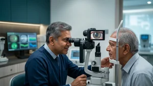
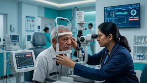
To reach a pigmentary glaucoma diagnosis it requires a full eye examination rather than a single test. An ophthalmologist will look for evidence of pigment dispersion along with any measurable effect on the optic nerve.
| Eye Test | Why It May Be Performed |
| Eye pressure measurement | Measures intraocular pressure and identifies levels that are elevated or fluctuating |
| Slit lamp examination | Allows the ophthalmologist to look for pigment deposits and changes in the iris |
| Gonioscopy | Examines the drainage angle and how much pigment has collected in the trabecular meshwork |
| Optic nerve examination or OCT | Assesses the optic nerve and retinal nerve fiber layer for signs of glaucoma related damage |
| Visual field test | Checks for areas of peripheral vision loss linked to glaucoma |
| Corneal thickness measurement | Helps the ophthalmologist interpret eye pressure readings more accurately |
How Is Pigmentary Glaucoma Treated?
The main objective of pigmentary glaucoma treatment is to lower your eye pressure sufficiently to minimize the chances of damaging your optic nerve. Your doctor will decide which treatments may be right for you based on factors such as your level of eye pressure, your examination findings related to your optic nerve, and results from tests performed on your visual field. It also considers things such as your age and how quickly your disease seems to be advancing.
Your doctor will likely start by prescribing eye drops that decrease how much fluid the eye makes or help your eye drain more effectively. It’s crucial that you follow your medication regimen carefully because glaucoma treatment focuses on providing long-term management, and if you stop your medications without proper instructions from your doctor, your eye pressure can increase.
Some people with pigmentary glaucoma symptoms ask about laser treatment as an option within their treatment plan. Selective laser trabeculoplasty (SLT) does target the trabecular meshwork specifically, however, because there is typically quite a bit of pigment loaded into the drainage angle [of someone’s eye] if they have pigmentary glaucoma, we try to determine whether this would be appropriate. Which also suggests your ophthalmologist to figure out what specific procedure makes sense for you based on the unique anatomy of your eye.
Medication and laser treatment combined aren’t always sufficient to manage someone’s intraocular pressure. That’s when a conversation can turn toward surgical options for managing pigmentary glaucoma. Traditional glaucoma surgery, a drainage implant, or a newer minimally invasive glaucoma procedure could be suggested by your ophthalmologist depending on how severe the problem is and other factors affecting your eye. All of these procedures are designed to help create a better pathway through which your eye can get rid of its extra fluids, thus alleviating the pressure.
Conclusion
This form of the disease arises when pigment from your iris interferes with the eye’s drainage system. It then puts the optic nerve in danger as the process continues. Because there are often no symptoms during the early phases, this disease tends to go unnoticed until you notice that vision has changed by which time, there could be irreversible damage already done.
Only an extensive evaluation (including measuring your eye’s pressure, performing a procedure called “gonioscopy” evaluating your optic nerve and using OCT imaging along with visual field testing) will give us enough information to tell if you’re suffering from just pigment dispersion syndrome, or full-blown pigmentary glaucoma. If you do end up having glaucoma, it needs to be treated based on what’s found during the evaluations.
Frequently Asked Questions
1. Is pigment dispersion syndrome the same as pigmentary glaucoma?
No. Pigment dispersion syndrome refers to pigment release from the back of the iris into the eye. Pigmentary glaucoma is diagnosed if this is accompanied by optic nerve damage (usually along with high/variable eye pressure).
2. What ages do people generally develop pigmentary glaucoma at?
The condition can affect younger and middle-aged adults and typically develops before most other types of glaucoma. However, age isn’t the sole determining factor for whether you’ll develop this condition. If you have unusual eye pressure measurements, find anything off about your optic nerves, or have a history of pigment dispersion, it’s crucial that you follow the exams recommended by your ophthalmologist.
3. Does exercise cause an increase in eye pressure if you suffer from pigmentary glaucoma?
Some people suffering from pigment disperson syndrome are prone to experiencing pigment release and temporary increases in eye pressure when they engage in very intense workouts. But that doesn’t mean that these patients should stop doing physical activities. You could discuss your workout plan with your ophthalmologist instead of immediately halting all forms of physical exertion.
4. Can pigmentary glaucoma ever be permanently cured?
Unfortunately there’s no way yet to reverse optic nerve damage once glaucoma has done it. Pigmentary glaucoma treatment will however help you keep your eye pressures down and prevent further optic nerve damage from happening. Most people with pigmentary glaucoma require on-going, long term follow-up for life, even if they’re doing really well otherwise.
5. How often does someone with pigment dispersion need to have an eye exam?
Patients who are diagnosed with stable pigment dispersion syndrome may require different monitoring than those who’ve been diagnosed with pigmentary glaucoma symptoms, which usually indicates more severe vision loss. Your ophthalmologist will be able to put together an exam schedule that’s tailored to your unique risks.