
An atypical look to the optic nerve may cause worry during an eye exam. One reason is optic disc drusen, which are small deposits that form in the optic nerve head. We see the condition put up by doctors during a routine eye health check. Also, people optic disc drusen may have the former which in turn causes damage to peripheral vision or in which the nerve appears to be raised, which in turn may present as if the optic disc swelling that is why a thorough work up is very important.
The optic nerve is what transmits visual info to the brain which is why docs pay atypical signs attention. They have to determine if a swollen looking nerve is due to drusen or some other issue. Some optic nerve disorders require prompt investigation, others a watch and wait approach.
What Are Optic Disc Drusen?
Optic disc drusen are calcified deposits in the optic nerve head. MedlinePlus reports that optic nerve head drusen are small reservoirs of protein and calcium that build up over time. These are different from retinal drusen in macular degeneration.
Some deposits are at the surface, in other cases they are buried. Buried deposits may cause the optic disc swelling to appear elevated and also present .We use imaging to determine the cause.
Many deposits are stable, in some patients we see visual field changes. We use monitoring to track this eye nerve disease and identify what is meaningful.
Why the Condition Can Look Like Swelling
Distinguishing between optic disc drusen and optic disc swelling is a key part of diagnosis. True swelling may be due to inflammation, decreased blood flow, compression, or raised pressure in the brain. Papilledema is a term we use for optic disc edema caused by raised intracranial pressure.
Drusen also causes pseudopapilledema which is a condition in which the nerve head appears raised without the presence of true edema. This distinction is important because some eye conditions that affect the optic nerve disorders may lead to loss of vision or be a sign of a neurologic
| Feature | Drusen | True Disc Swelling |
| Main issue | Deposits within the optic nerve head | Genuine edema from an ocular or neurological cause |
| Symptoms | Often absent; field changes can occur | Depend on the underlying cause |
| Appearance | Disc may look elevated or irregular | Disc may appear elevated with signs of edema |
| Evaluation | Eye examination and targeted imaging | Eye examination plus investigation of the cause |
| Usual approach | Monitoring after diagnosis | Treatment or urgent assessment may be needed |
Common Symptoms
Most of what we see in this condition is asymptomatic patients. Central visual acuity may be preserved. What changes do appear tend to affect the peripheral visual field. Also some may report brief episodes of vision which are dim or vague.
Small field issues may go by unsaid in daily life. We are able to detect them with visual field testing. Also symptoms present with other optic nerve disorders which makes self diagnosis out of the question.
Sudden vision loss, notice that which is a new blind spot, severe headache, double vision, or very rapid changes in what you see should be looked at quickly by a professional. Also an eye nerve disease or what is known as optic disc swelling may be present.
Why Do These Deposits Develop?
The exact cause is out to determine, as with many optic nerve disorders. Present medical research reports that drusen formation is associated with changes in axonal metabolism and the movement of material within optic nerve fibers. Calcified material may also accumulate in the nerve head.
Anatomy does play a role, also we see that there are familial trends. Screen use, reading, and normal eye habits don’t account for it.
A thorough evaluation differentiates our findings from other optic nerve disorders, which may have different risk factors and treatment options.
How Are Optic Disc Drusen Diagnosed?
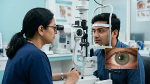
An ophthalmologist will first do a look at the optic nerve and also to assess visual function. Superficial optic disc drusen may present during a dilated eye exam. Buried deposits which may be at first thought to be a case of optic disc swelling require imaging.
OCT provides transverse images of the optic nerve head and retinal nerve fibre layer. In Enhanced-depth OCT we are able to see through to greater depths. Autofluorescence may also present superficial drusen which B scan ultrasound may in turn identify in terms of calcification.
Visual field study which assesses side vision and documents functional change. Doctors may order it when symptoms or field defects are present.
| Test | What It Helps Assess |
| Dilated eye examination | Optic nerve appearance and visible deposits |
| OCT | Optic nerve head and retinal nerve fibre layer |
| Enhanced-depth OCT | Deeper or buried deposits |
| Fundus autofluorescence | Superficial deposits |
| B-scan ultrasound | Calcified deposits in selected cases |
| Visual field test | Peripheral vision and functional change |
If what is found does not clearly point to optic disc drusen the doctor will look at other optic nerve disorders. This is particularly true when symptoms point towards true optic disc swelling or another eye nerve disease.
Is the Condition Dangerous?
In many people the issue doesn’t produce major central vision loss. That said, the issue shouldn’t be ignored. Visual field defects do report in some and also very rarely we see vascular complications related to the optic nerve.
Risk varies between individuals. In some cases repeat evaluation is enough, but in others which present with variable visual fields more frequent follow up is required. Also we do not want to immediate attribute every new symptom to an existing eye nerve disease.
Treatment and Monitoring
There is at present no sure treatment for optic disc drusen or which in all cases will prevent related field loss. What we see is that which reports are few and that care is mostly about diagnosis and monitoring of vision and nerve structure.
Follow up will include visual acuity, eye pressure, OCT, and visual field testing. Timing of the follow up will depend on symptoms, findings, age, and change over time.
If there is a different issue which develops we treat that which is at the root of it. Drusen may present with another eye condition at the same time which is of the eye nerve disease type, in that case should have the optic disc swelling be assessed.
When Should You See an Eye Specialist?
Arrange for an eye exam if your optic nerve appears raised, puffy, or out of the ordinary. Also report in on unexplained blind spots, recurring dim vision or changes in peripheral vision.
Urgency in evaluation is of great importance when one presents with sudden visual issues which also include severe headache, vomiting, double vision, weakness, or confusion. Severe optic nerve disorders may present with these.
For patients which have already been diagnosed we see that regular follow up is a help to note if the condition is stable. We also note that from previous OCT and visual field reports we have useful comparisons to make when reviewing this eye nerve disease.
Living With Optic Disc Drusen
Many patients who have optic disc drusen do well and preserve useful central vision. Regular follow up is important for this eye nerve disease.
Tell new eye care professionals of the diagnosis. It may help them to recognize an enlarged looking disc but also to confirm or rule out true optic disc swelling.
At ASG Eye Care we use optic nerve imaging and visual field testing to determine the cause of an atypical disc appearance and to guide our monitoring of eye nerve disease.
Conclusion
Optic disc drusen are collections of material at the optic nerve head which in many cases produce few symptoms. They have a raised appearance which in turn may resemble optic disc swelling which is why accurate diagnosis is key. OCT, visual field testing, autofluorescence, and ultrasound may be used to tell drusen apart from other eye nerve disease.
Most of the time we see that patients require a period of observation. We do routine checks which also serve to document any change. At the first sign of acute visual or neurological symptoms get in to see the doctor. We recommend you seek prompt care because some problems with the optic nerve disorders are a medical emergency.
Frequently Asked Questions
1. Can optic disc drusen cause blindness?
Optic disc drusen generally does not lead to total blindness. Some people do develop peripheral visual field loss, but central vision may remain relatively preserved. Regular testing is for tracking changes.
2. Are optic disc drusen the same as papilledema?
No. Drusen may present as pseudopapilledema which is a mimic of optic disc swelling. Papilledema is real swelling from raised intracranial pressure. It is important for doctors to tell the difference between the two which has different medical implications.
3. Can children have optic disc drusen?
Yes. Optic disc drusen do present in pediatric patients and the deposits may be more internal within the nerve head. Diagnosis may be confirmed via imaging which is useful when the presentation is atypical.
4. Do optic disc drusen need surgery?
In most cases surgery is not required. We do not have a standard procedure which will remove the deposits. Care typically involves monitoring and reporting of complications as well as other optic nerve disorders.
5. Can the condition get worse?
The course of the disease may also present with changes in appearance over time and in some patients visual field defects progress. Routine follow up is important for comparison of nerve structure and function which in turn may identify the onset of eye nerve disease.
6. Can it occur with another optic nerve problem?
Yes. Optic disc drusen may present with some other optic nerve disorders. Presenting symptoms should be reevaluated which is to say don’t assume they are a result of the current diagnosis.