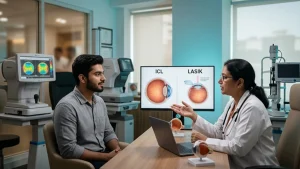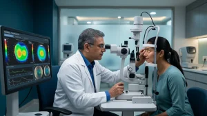
LASIK does in fact reduce the use of glasses or contact lenses. But not all eyes are a good fit for laser vision correction. Prior to putting a patient under for the procedure an ophthalmologist will do an exam of the cornea which is the clear front of the eye. We do corneal mapping before LASIK which gives us info on it’s shape, curvature and thickness. That info in turn we use to create a better treatment plan which is safer.
In contrast to a regular vision test which is routine in nature, corneal topography which we use is a in depth evaluation of the cornea. Also it brings to notice irregularities which may not be a part of what a standard eye exam includes. Also we use this info for keratoconus screening and to determine LASIK eligibility. By understanding these tests patients are better able to make informed decisions regarding their vision.
What is Corneal Mapping pre-LASIK?
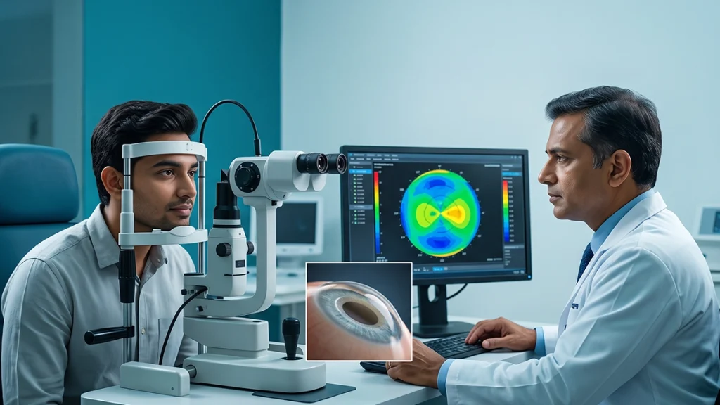
corneal mapping before LASIK is a diagnostic test which looks at the shape and surface curvature of the cornea. Specialized equipment takes in the measurements which it then puts into a color coded map. Different colors represent different levels of curvature.
This procedure is without pain, does not require any invasion, and typically takes a few minutes. Patients will look at a target which the machine will use to take images. The results of which help doctors to determine the eye’s natural form which in turn will inform treatment.
During corneal mapping before LASIK the specialist also may order extra imaging which studies the cornea’s thickness and internal structure. This info gives a full picture of corneal health.
What does Corneal Topography show?
This test identifies which areas of the Corneal topography are steep, flat, and irregular. These patterns in turn explain some cases of blurry vision and astigmatism. Also it helps surgeons to determine how the cornea will respond to laser procedures.
The following table explains what doctors may assess during corneal mapping before LASIK.
| Finding | What It May Reveal |
| Corneal curvature | Steep or flat areas affecting focus |
| Surface irregularities | Uneven curvature or unusual patterns |
| Corneal thickness | Whether sufficient tissue may remain after treatment |
| Asymmetrical patterns | Possible signs of an underlying corneal condition |
Corneal topography does present with atypical patterns which may raise red flags but it is not to be used as a stand alone diagnostic tool. Doctors put these into the context of other test results.
Why Keratoconus Screening Matters
Keratoconus is a condition that the cornea which is to become thinner over time does in fact bulge out. This causes changes which at the end cause distortion of vision and increased astigmatism. In early stages symptoms may not be at all present.
Keratoconus screening is used by doctors to identify early signs of corneal degeneration before they may recommend surgical options. That is important to note as the various laser procedures that we have are designed to alter the cornea’s surface. To put on an eye which is unknown to have cornea weakness at risk is to increase the chance of issues.
corneal mapping before LASIK may present patterns that indicate a more in depth look. At which time doctors may order corneal tomography or other diagnostic tests. A positive result doesn’t confirm keratoconus. That is determined by the whole clinical picture the specialist looks at.
People which present with a history of keratoconus in your family, getting new prescriptions, or which have unexplained blurred vision should bring that up at your eye exam.
How Mapping Helps Determine LASIK Eligibility
LASIK eligibility which is a type of vision correction procedure is not based only on a patient’s glasses prescription. The doctors will look at the corneal shape, the thickness, prescription stability, tear film health, and the overall eye health
Corneal mapping before LASIK allows the surgeon to determine if the cornea is a good candidate for the procedure. Should results indicate a high risk of complication the doctor may present alternative solutions or may put forward that LASIK is not the proper treatment.
| Assessment | Why It Matters |
| Corneal shape | Identifies suspicious or irregular patterns |
| Corneal thickness | Supports safe treatment planning |
| Prescription stability | Helps confirm that vision measurements remain consistent |
| Tear-film health | Reduces measurement errors caused by dry eyes |
| Overall eye health | Helps identify other factors affecting surgical suitability |
LASIK eligibility is on a case by case basis. No single measurement is a go or a no go for success
What To Expect Before the Test?
Your eye care professional will ask into your past health including past eye issues, current meds, and recent changes to your prescription. Contact lenses which may change the shape of the cornea are a factor, so you may be asked to go without them before the test. The number of days which to abstain depends on the type of contacts you wear and what we find in the initial exam.
During corneal mapping before LASIK try to follow the tech’s instructions and keep your eyes open as told. Report to the team if you have discomfort or have difficulty with the target.
The specialist will go over the results and also will see if more tests are required. Even a normally appearing map does not take the place of a thorough preop eye exam.
Conclusion
Corneal mapping before LASIK is a key tool in which we determine eye shape and also which we use to identify what issues may present during the procedure. It is a way for specialists to study the cornea, to look at irregularities, and to put forth informed treatment plans. Also it is made known that not all patients will be suitable for this procedure.
At ASG Eye Care our staff will go over your vision goals with you and also have you meet with an eye specialist for a preop evaluation. We do a very in depth assessment which in turn will inform you of all your options and help you to make that sure footed, informed decision on which vision correction is right for you.
Frequently Asked Questions
1. Is it painful for corneal mapping?
No. The test is non invasive and usually does not cause any discomfort. The machine takes in images of the cornea without touching it during the procedure.
2. Can corneal mapping detect keratoconus?
It will point out which elements of the picture don’t add up. Also in some cases we will need to do more tests to confirm the diagnosis and see the stage of the disease.
3. Does routine corneal mapping determine if a patient is a good candidate for LASIK?
No. Also they look at corneal thickness, prescription stability, dry eye issues and other health factors.
4. How long can you expect to be in the office for?
The actual imaging of the procedure only takes a few minutes. The full pre-LASIK work up may take more time which is due to extra tests.
Government and Medical Reference Links
- U.S. Food and Drug Administration (FDA): When Is LASIK Not for Me?
https://www.fda.gov/medical-devices/lasik/when-lasik-not-me - National Eye Institute (NEI): Corneal Conditions and Keratoconus Information
https://www.nei.nih.gov/eye-health-information/eye-conditions-and-diseases/corneal-conditions/corneal-dystrophies - U.S. Food and Drug Administration (FDA): LASIK Patient Information
https://www.fda.gov/medical-devices/surgery-devices/lasik