
Introduction
Blood, a dense cataract or some other obstruction may prevent the doctor from getting a clear view of the retina. A B scan eye test uses sound waves to produce images in cases which do not provide enough detail in a routine exam.
What Is a B-Scan Ultrasound?
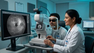
B scan eye test produces a 2D image of the inner structure of the eye. This test is used by doctors when a clear view of the eye is not present. The B scan eye test sends out sound waves which return images of what the waves echo. Also known as ophthalmic ocular ultrasound it is a tool which doctors use when they are unable to see in to the eye.
Doctors will order this scan in a case where they are unable to do a routine exam of the retina. That which may present in vitreous bleed, a dense cataract, or with some injuries. The ophthalmologist will put the results of the scan in context with symptoms and examination findings.
When Might a Doctor Recommend It?
B scan eye test is a tool we use when there is blood in the vitreous gel which in turn is blocking the view of the retina. This is what we term as a vitreous hemorrhage. We use the vitreous hemorrhage scan to help us identify other issues within the retina which may be out of sight, also to look for a possible tear or detachment.
If a doctor thinks there is retinal detachment but is unable to see the retina clearly a retinal detachment ultrasound may help in diagnosis. The results of aB scan eye test should be put in the context of other reports.
What Can the Scan Help Detect?
B scan eye test is used to investigate many issues which the retina does not allow for proper in person assessment
| Condition | How ultrasound may help |
| Vitreous haemorrhage | A vitreous hemorrhage scan may help assess the retina when blood blocks the view. |
| Suspected retinal detachment | A retinal detachment ultrasound may reveal separation of the retina. |
| Dense cataract | An ocular ultrasound can help examine structures behind a cloudy lens. |
| Eye injury | Ultrasound may reveal internal abnormalities. |
A vitreous hemorrhage scan may present an issue at times when blood is between the lens and retina that in turn affects image quality and what we see. Retinal detachment ultrasound will give us that extra info which we put into practice; in the end the ophthalmologist will make the diagnosis.
How Is the Test Performed?
B scan eye test does not require much preparation. The patient sits or lies down and the doctor applies gel to the closed eye. The doctor then moves a probe over the eyelid which in turn captures images.
The procedure is free of pain and we do not use radiation. Before we do the test tell us if there has been any recent injury or discomfort.
| Test detail | What to expect |
| Preparation | Usually no special preparation is needed. |
| Method | A probe moves gently over the closed eyelid with gel. |
| Comfort | The procedure is generally painless. |
| After the scan | The ophthalmologist explains the results. |
Why Do We Need to Seek Immediate Care for Retinal Detachment?
A retinal detachment ultrasound is used when the doctor has reason to think there is a detachment but is not able to see the retina with the naked eye. Retinal detachment is a severe issue which may lead to permanent vision loss if treated late. Signs like sudden flashes, large numbers of new floaters, a dark curtain that comes into your field of vision, or a sudden change in your vision should be brought to the doctors’ attention right away.
A vitreous hemorrhage scan may be diagnosed with a scan which also looks at related retinal issues. Should you see warning signs do not wait for the scan appointment. The ophthalmologist uses results of the B scan eye test to determine the next steps.
Conclusion
B scan eye test is used by ophthalmologists to assess the back of the eye in cases of blood, a cataract, or other interference which causes a poor view. Also we have ocular ultrasound, vitreous hemorrhage scan, or retinal detachment ultrasound which support in this evaluation. For sudden flashes, new floaters, a curtain like shadow, or sudden vision loss seek immediate eye care.
Frequently Asked Questions
1. Is a B-scan ultrasound painful?
B scan eye test is a pain free procedure. We apply gel to the closed eye and gently move the probe. Report any recent eye injury or pain before the procedure.
2. Ultrasound also may be used to detect retinal detachment ?
A retinal detachment ultrasound is a tool which helps in the diagnosis of retinal separation when the eye does not allow a clear view of the retina. The ophthalmologist will determine if treatment is urgent.
3. Why do I need a vitreous hemorrhage scan?
A vitreous hemorrhage scan may be recommended when you have blood in the eye which is blocking the view of the retina. It also helps the doctor to see if there is a tear or detachment.
4. Is an ocular ultrasound the same as a regular eye exam?
No. In an ocular ultrasound we use sound waves to produce internal images. In a regular exam we do vision and retinal testing. Doctor will perform tests based on symptoms and if an ocular ultrasound is required
Reference Links
1. National Eye Institute: Retinal Detachment –
https://www.nei.nih.gov/eye-health-information/eye-conditions-and-diseases/retinal-detachment