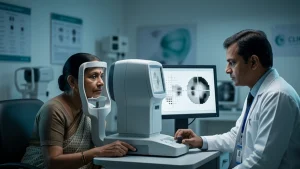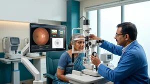
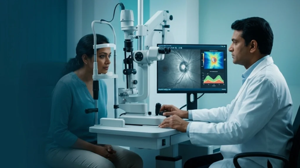
Glaucoma may also slowly impair your vision with some people experiencing no obvious symptoms in which time the issue may go unnoticed. What does change is that during it\’s early stages we do not always see it, that is why regular eye exams are very useful in which they are able to spot things before you will. Also a key test used is the OCT scan which in detail maps out the structures within your eye.
An RNFL OCT glaucoma does what ophthalmologists do which is look at the nerves at the back of the eye. These are the fibers which take visual info from the retina to the brain. We use this to see changes in their thickness which in turn helps us to identify glaucoma related changes and also to see how the disease progresses over time.
What Is an RNFL OCT Scan?
RNFL which is Retinal Nerve Fibre Layer. This layer is made up of nerve fibers which are a part of the optic nerve. In glaucoma these fibers are damaged which in turn cause the RNFL to thin out.
An OCT scan uses light waves to produce in depth cross sectional images of the eye. Also unlike an X-ray it does not use radiation. The test is non-invasive and usually takes only a few minutes.
An RNFL OCT glaucoma assessment measures the thickness of the nerve fiber layer and compares it to a reference database. Also, your ophthalmologist may look at the optic nerve and take into account your eye pressure and visual field results.
How Does OCT Help Track Glaucoma?
An OCT optic nerve provides doctors with a detailed look at the nerve and the retina which surrounds it. It is also able to identify structural changes that may not yet be causing visual symptoms.
Doctors also use these scans as a base for future comparison. If we see progression in the repeat scans, our ophthalmologist will look into whether the glaucoma is worsening. That is to say, RNFL OCT glaucoma testing is very useful for long-term monitoring.
However in a single scan not all cases will be diagnosed. Doctors look at the results in conjunction with your medical history, physical exam results and other glaucoma tests.
What Do RNFL OCT Results Mean?
OCT reports that colors are used to compare present results with a reference database. Green is to indicate that which is within the normal range, while yellow or red is used for thinner measurements.
These results do not present a diagnosis by themselves. Some healthy eyes may have thinner nerve fibre layers. Also other eye conditions, scan quality and individual anatomy play a role in results.
Table 1: Understanding Common OCT Report Findings
| OCT finding | What it may indicate |
| RNFL thickness within the expected range | No clear thinning in the measured areas |
| Reduced RNFL thickness | Possible nerve fibre loss that needs clinical interpretation |
| Asymmetry between the eyes | A difference that may require further assessment |
| Progressive thinning on repeat scans | Possible structural change associated with glaucoma |
Your eye doctor will go over your results with you. A color coded report does not in and of itself determine if you have glaucoma.
Can RNFL OCT Detect Glaucoma Progression?
An RNFL OCT glaucoma which may be used by doctors to identify structural changes between office visits. Progressive thinning is an indicator of glaucoma progression which in the case of the same between other assessment results’ reports points very much to that which is diagnosed.
Doctors, which is what we do we put present scans against past results instead of reporting off of a single measurement. Also we look at the reliability of the scans and we see if other eye conditions play a role in what we are seeing.
Table 2: OCT Monitoring During Glaucoma Care
| Monitoring stage | Purpose |
| Initial OCT scan | Establishes a baseline for comparison |
| Follow-up OCT scans | Helps identify changes in nerve fibre thickness |
| Visual field testing | Checks for changes in peripheral vision |
| Comprehensive eye examination | Combines scan results with pressure and optic nerve findings |
OCT and visual field testing reports which have distinct sets of data. The former looks at structure changes and the latter at function within your field of vision. Thus together they play a role in which doctors base their diagnosis on.
Who May Need an RNFL OCT Scan?
Your ophthalmologist may suggest an RNFL OCT for glaucoma if you have what is thought to be glaucoma, a history of the disease in your family or changes in your optic nerve. We also use OCT to track the progress of glaucoma in people that already have it.
The testing schedule we recommend is based on your diagnosis, past results and overall eye health. Very important to note that early glaucoma may not present with noticeable symptoms.
Conclusion
An RNFL OCT glaucoma scan is used by ophthalmologists to monitor health of the optic nerve and to identify changes which may go undetected. When doctors perform comparisons of scans over time they are able to better assess disease progression and to determine if your treatment plan needs to be changed.
At ASG Eye Care we offer in depth eye exams which will help you to understand your results and what the next steps are for protecting your vision. If you have glaucoma or are worried about the health of your optic nerve see an eye specialist and follow the recommended monitoring schedule.
Frequently Asked Questions
1. Is an RNFL OCT scan painful?
No. An RNFL OCT scan is a non-invasive and usually painless procedure. All you do is look at a target as the machine takes images of your eye. This RNFL OCT glaucoma which also helps to determine the health of your optic nerve.
2. Can OCT confirm glaucoma on its own?
No. Doctors also use a combination of tests which may include ophthalmoscopic views of the optic nerve, measurement of eye pressure and visual field testing. An OCT optic nerve scan is a tool which doctors use to see structural changes which may be early indicators of glaucoma.
3. Does a normal OCT result mean I cannot have glaucoma?
Some of the time. We see that some people do in fact have glaucoma related changes which may require more in depth testing even when the OCT results look normal. Evaluation of the retinal nerve fiber layer may reveal structural changes that may not be apparent in a basic vision assessment.
4. How often should I get an OCT scan?
Your eye doctor will create a schedule for you based on your condition, risk factors and past results. Some patients require more frequent RNFL OCT glaucoma follow up than others, especially when early scans show change in the retinal nerve fibre layer.
5. Can OCT show whether glaucoma is getting worse?
Repeat scans which over time will show structural changes. Your doctor will compare these results with visual field results and other clinical assessments to evaluate possible glaucoma progression. Regular RNFL and OCT optic nerve scans for glaucoma also help monitor changes and make informed decisions about your care.
References link
National Eye Institute — Glaucoma: Symptoms, Diagnosis and Treatment
https://www.nei.nih.gov/eye-health-information/eye-conditions-and-diseases/glaucoma
UK National Health Service — Glaucoma Imaging Clinic: OCT and Monitoring
https://leaflets.ekhuft.nhs.uk/glaucoma-imaging-clinic/html/
UK National Health Service — Glaucoma Monitoring Tests
https://www.dgft.nhs.uk/pil/glaucoma-and-monitoring-tests/