Eye issues may not present with clear signs of vision change. At times the eye will show a health issue which is from somewhere else in the body. . Papilledema is an example of this. It is a condition which consists of swelling of the optic disc which in turn is a result of increased pressure within the skull. The optic disc is that area where the optic nerve meets the back of the eye and is what transmits visual info to the brain.
Because of the association between papilledema and raised intracranial pressure we take it very seriously. The cause may range from idiopathic intracranial hypertension to conditions which require acute medical attention. Early evaluation is key in identifying the cause and in protecting the optic nerve from permanent damage.
According to the U.S. National Library of Medicine’s NCBI medical report which details papilledema the condition results from raised intracranial pressure which in turn causes swelling of the optic disc. Also reported is the fact that it is important to tell it apart from other types of optic disc swelling.
What Is Papilledema?
Papilledema is a condition of optic nerve head swelling which is a result of increased intracranial pressure. It usually affects both eyes, though the swelling may not present equally in each eye. Other diseases may cause a puffy optic disc. But we use this term in particular when brain pressure is the cause of the swelling.
The pressure which in turn interferes with normal flow within optic nerve fibers. As swelling progresses the optic disc becomes raised and its edges less distinct. If left untreated this may lead to damage of the optic nerve fibers and in turn affect vision.
A person may present with papilledema before they notice any large scale changes in vision. That is why a very detailed eye exam is of great importance which in turn we should perform especially when there are headaches or other neuro symptoms associated with visual disturbances.
What Causes Papilledema?
In many health issues the pressure in the skull may increase which in turn may cause papilledema. A very common cause is Idiopathic Intracranial Hypertension. In this disease the intracranial pressure goes up without a mass in the brain being found.
Other possible causes are a brain tumor, brain abscess, intracranial bleeding, head injury, meningitis, encephalitis and obstructive hydrocephalus. Cerebral venous sinus thrombosis also causes increased intracranial pressure.
This large spectrum of causes which we see plays that doctors do not treat optic disc swelling as a stand alone eye issue. Identifying what is causing the pressure to increase is a very important part of papilledema treatment.
Common Papilledema Symptoms
At the onset papilledema symptoms may present with very mild symptoms. Some report no change in visual acuity at all in the early stage. Others note what are called transient visual obscuration in which the vision may go gray, blurred, or dark for a few seconds. These events at times are referred to as flashers.
Headache is a very present symptom which in turn becomes more notable when intracranial pressure has increased. Also patients may report nausea, vomiting, double vision or a pulse like whooshing sound in the ears. The pattern and degree of papilledema symptoms varies with the base cause and the level of pressure.
As the papilledema advances peripheral vision may be affected. Severe or long term disease at some point may put central vision in jeopardy because of ongoing pressure which in turn damages the optic nerve.
| Possible Symptom | What a Patient May Notice |
| Headache | Persistent or recurring head pain |
| Temporary visual disturbance | Brief dimming, greying or blurring of vision |
| Double vision | Two images instead of one |
| Pulsatile tinnitus | A rhythmic whooshing sound in the ears |
| Nausea or vomiting | Often associated with increased intracranial pressure |
| Visual field changes | Reduced peripheral vision in more advanced disease |
Stages of Papilledema and the Frisén Scale
Doctors may use a scale which they have designed which to stages of papilledema based on what they see in the optic disc. The most used is the modified Frisén scale which grades the appearance of the optic disc from 0 to 5. 0 grade indicates a normal disc and 5 a very severe swelling.
The grade of papilledema is a tool which clinicians use to report out the degree of swelling which is present and to track changes over time. But that grade does not tell us the cause. A person still requires appropriate investigation to determine what is behind the increase in intracranial pressure.
Understanding Papilledema Grading
The following table provides a simplified overview of papilledema grading based on the Frisén system. Clinical assessment involves more detail than this summary.
| Grade | General Appearance |
| Grade 0 | Normal optic disc without true swelling |
| Grade 1 | Minimal swelling with early changes around the disc margin |
| Grade 2 | Circumferential halo and clearer evidence of early swelling |
| Grade 3 | Moderate swelling with obscuration of segments of major vessels |
| Grade 4 | Marked swelling with greater vessel obscuration and disc elevation |
| Grade 5 | Severe swelling with extensive obscuration of vessels on and leaving the disc |
The progression of stages of papilledema may present a structured approach to documentation. A higher grade of papilledema usually means more visible optic disc swelling. In grade 3 we see moderate swelling and which in turn blinds out one or more large vessel segments. Grades 4 and 5 present more severe changes.
Doctors may include in their assessment of papilledema grading the use of visual field testing, optic nerve imaging and other studies. This approach gives a more complete picture of the health of the optic nerve as opposed to based only on appearance.
How Is Papilledema Diagnosed?
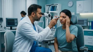
Diagnosis of papilledema begins with a medical history and eye exam. An ophthalmologist looks at the optic disc for signs of swelling, blurred edges, modified blood vessels and other key indicators.
Visual acuity and visual field tests may also be performed. Visual fields in particular are useful which is because an enlarged blind spot or peripheral field defect may appear even as central vision stays relatively unaffected.
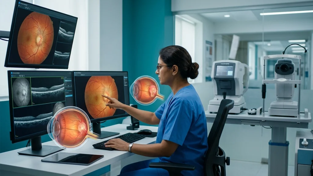
OCT is what we also term as Optical Coherence Tomography which measures retinal nerve fiber layer thickness. Doctors use OCT to document swelling and to monitor changes over time. But it is important to interpret OCT results in the context of the full clinical picture.
When papilledema is reported we go beyond ophthalmic evaluation. Brain imaging which is usually an MRI or in some cases a CT is ordered to look for a mass in the brain or any other cause of increased pressure. MR venography or CT venography is used to study the cerebral venous system when it is relevant to the clinical picture.
After we have ruled out through imaging that a patient has a condition which makes the procedure unsafe, doctors may put forth a lumbar puncture. It is a tool which measures cerebrospinal fluid opening pressure and also allows for laboratory analysis of the fluid. These tests in turn help doctors determine proper papilledema treatment.
How Is Papilledema Treated?
There is not a universal papilledema treatment which we must bear in that it is a sign of a different health issue. What we do is focused on reducing intracranial pressure and treating the root cause.
For instance what we see is that the care provided varies greatly when pressure is a result of infection, bleeding, a mass, venous sinus thrombosis or Idiopathic Intracranial Hypertension. This is why we advise patients to try to treat their symptoms out which they may think is what is causing the issue.
There is not a universal papilledema treatment which we must bear in that it is a sign of a different health issue. What we do is focused on reducing intracranial pressure and treating the root cause.
For instance what we see is that the care provided varies greatly when pressure is a result of infection, bleeding, a mass, venous sinus thrombosis or Idiopathic Intracranial Hypertension. This is why we advise patients to try to treat their symptoms out which they may think is what is causing the issue.
Can Papilledema Be Prevented?
It does not always happen that we are able to prevent papilledema which is a result of what causes the raise in intracranial pressure. Some base conditions which cause this may present itself unexpectedly and require medical attention instead of a routine preventive approach.
The early sign of which to look out for is that of warning signs and we recommend you seek assessment for them instead of putting forth that you ignore persistent headaches or new visual disturbances. Also for people that are already in care and treatment for a condition which has raised intracranial pressure we ask that they follow their prescribed care and monitoring plan.
Regular follow up also allows doctors to note changes in the grade of papilledema, optic nerve appearance and visual function over time. Early detection of disease worsening may which in turn permit a change in treatment before permanent visual damage occurs.
When Should You Consult an Eye Specialist?
New visual symptoms should not be put on hold when they present with severe and persistent headache, nausea, vomiting, double vision or pulsatile tinnitus. Acute or progressive loss of vision requires prompt medical evaluation.
An ophthalmologist can identify if disc bulging is due to papilledema or some other issue. Should there be a case of raised intracranial pressure we may put forward for a neurological evaluation and imaging immediately if it is urgent not to wait.
Recognizing papilledema symptoms at an early stage is very important as eye sight may still be normal in the early stages. By the time major vision loss occurs the disease may already be in an advanced stage which in turn delays diagnosis.
Conclusion
Papilledema is not just a case of what one sees at an ophthalmic exam. This is an important indicator which in fact reports raised pressure within the skull. While sight may be to a large degree unaffected in the initial stages, in the long term it will produce damage to the optic nerve.
Understanding papilledema symptoms, its stages of papilledema and the role of papilledema grading will help patients see the value in timely investigation. The right treatment for papilledema is to identify and manage the cause of increased intracranial pressure.
If you experience headaches in association with new visual changes, double vision, temporary vision loss or any other serious symptoms see a doctor right away. An ophthalmologist may perform an exam of the optic nerve, study visual function and also order more tests when there is a suspicion of increased intracranial pressure.
Frequently Asked Questions
1. What is the main cause of papilledema?
Papilledema is a result of increased pressure in the skull. Causes may include idiopathic intracranial hypertension, brain masses, bleeding, infections, hydrocephalus and cerebral venous sinus thrombosis. It is important that the root cause is determined before treatment is applied.
2. What are the early papilledema symptoms?
Early papilledema symptoms may present as headache, temporary blurred or dim vision, brief visual blackouts, double vision and pulsatile tinnitus. Some people may have only mild visual symptoms at first which is the reason why an eye exam plays a key diagnostic role.
3. What are the stages of papilledema?
The stages of papilledema are typically done via the Frisén scale. This goes from Grade 0 which is a normal optic disc to Grade 5 which is severe swelling that also includes extensive blood vessel obstruction.
4. What does the grade of papilledema mean?
The grade of papilledema is a term which we use to describe the degree of optic disc swelling. Doctors look at features like disc proptosis, unclear borders and compression of retinal vessels. Grading of papilledema is used to document the degree of severity and to track changes over time but it does not identify the cause.
5. How does papilledema grading help doctors?
Papilledema grading is a standardized method of reporting optic disk changes at exam. Also by using grades which correspond to OCT results and visual field studies doctors may track the course of the disease and determine if there is improvement or deterioration of the edema.
6. Can papilledema cause permanent vision loss?
Yes. Long term or severe papilledema may cause damage to the optic nerves which in turn leads to permanent visual damage. Timely investigation and treatment of raised intracranial pressure is a way to reduce that risk of progression of optic nerve damage.
7. What tests are used to diagnose papilledema?
Doctors may carry out a dilated eye exam, visual field testing and OCT. In the case of raised intracranial pressure we usually see imaging of the brain. Also a lumbar puncture may be done when it is determined by the clinician to be safe and medically appropriate.
8. Is papilledema treatment always surgical?
No. Papilledema treatment is based on the cause, severity and risk to vision. In some cases we may do medical management and in others we may go for procedures or surgery which will help to control the pressure or to treat the base cause.
Reference links
- Papilledema – NCBI Bookshelf, U.S. National Library of Medicine (NIH)
https://www.ncbi.nlm.nih.gov/books/NBK538295/?utm_source=chatgpt.com - Papilledema – PubMed, U.S. National Library of Medicine (NIH)
https://pubmed.ncbi.nlm.nih.gov/30855879/?utm_source=chatgpt.com - Pseudopapilledema – NCBI Bookshelf, U.S. National Library of Medicine (NIH)
https://www.ncbi.nlm.nih.gov/books/NBK538291/?utm_source=chatgpt.com