
Changes in the iris color or shape may go unnoticed, also when visual acuity is the same at wake up, halos around lights or one sided eye pain may in fact point to this rare condition which affects the cornea, iris and the drainage angle of the eye.
This patient guide details the development of ICE syndrome, what symptoms to look for, the tests an ophthalmologist may order and the treatments which are put in place to protect sight. Although it is a rare eye disease, what you should look for is the warning signs which bring you in to see a specialist before permanent damage sets in. Also it is important to tell the difference between ICE and inherited corneal disorders and other causes of iris changes.
What Is Iridocorneal Endothelial Syndrome?
Iridocorneal endothelial syndrome is a group of eye diseases which see abnormally from the cornea’s endothelium spread to the drainage angle and the front of the iris. The corneal endothelium is the inner most layer of the cornea. It plays a role in pumping out extra fluid which in turn keeps that clear front window of the eye transparent.
When these atypical cells migrate they can form a membrane which over time may pull the pupil out of its normal position, thin out or damage the iris, close off parts of the drainage angle and interrupt the normal flow of fluid in the eye. This can cause increased eye pressure, glaucoma and corneal oedema. NIH reports visible iris changes, corneal swelling and glaucoma as the main features of the condition.
Inherited corneal disorders which present as Iridocorneal Endothelial Syndrome usually do so in adult life and typically affect one eye. It is reported more in women, especially in early to middle adulthood although men and those out of the usual age range also do develop it. Bilateral cases are atypical but have been reported.
The cause of iridocorneal endothelial syndrome is at present unidentified. What we do know is that researchers have looked into if inflammation or past viral triggers play a role in transforming corneal endothelial cells to take on properties of epithelial cells. Herpes simplex virus DNA was found in some of the tissue samples we looked at which is why we say that it may be a factor in some cases but we can’t yet say that it is the cause in all.
At present the condition is out to be of a spontaneous nature as opposed to a clear cut inherited disorder. As this rare eye disease is usually acquired which means it isn’t a genetic issue for the majority there is no reason to screen other family members once one case is reported. Main Forms Doctors also report that what we have termed iridocorneal endothelial syndrome is actually of three related forms. A patient may present with signs of more than one form also the presentation can change as the disease progresses.
| Form | Common clinical pattern | Possible effect on vision |
| Essential or progressive iris atrophy | Marked iris thinning, displaced pupil and sometimes full-thickness iris holes | Glare, distorted pupil appearance and a substantial risk of secondary glaucoma |
| Chandler syndrome | Corneal swelling tends to be more prominent, while iris changes may initially be subtle | Blurred vision, coloured halos and morning haze caused by corneal oedema |
| Cogan–Reese syndrome | Pigmented spots, nodules or a diffuse nevus-like appearance on the iris | Variable blur, cosmetic change, pressure rise and progressive glaucoma |
Chandler syndrome is noted to produce greater corneal oedema; also in some cases there is more evident iris distortion present in progressive iris atrophy. Cogan and Reese’s syndrome can appear as a pigmented iris lesion. We use these differences as a guide in our examination protocol, but it is important to know that all three of these conditions stem from the same defective endothelial process and may lead to glaucoma or corneal decompensation.
Like in some corneal diseases which present as a range of subtypes that affect corneal transparency these may also seem to present with changes in the iris and drainage angle which in turn present as very useful in diagnosis.
What Need Attention
The course of Iridocorneal Endothelial Syndrome is very variable. In some cases we see that one pupil has taken on an oval, displaced or irregular shape. In others the issue is of morning vision which is foggy which improves by the afternoon. Morning blur may be due to the cornea’s increased fluid content which happens while the eyes are closed at night.
Common symptoms are fluctuating blurred vision, halos around lights, sensitivity to glare, a change in iris color or pupil position, redness, headache and a pain around one eye. Sudden or severe pain, nausea, marked redness and quick loss of vision may be a sign of a great rise in eye pressure and requires prompt ophthalmic care.
This rare eye disease may present in a routine eye exam before the patient notes any primary symptoms. Its early stages may present like glaucoma, uveitis and other corneal diseases which makes self diagnosis unreliable.
| Finding noticed by the patient or doctor | What it may indicate | Recommended response |
| Morning haze or halos | Corneal oedema caused by weak endothelial pumping | Arrange an eye examination, especially if symptoms repeatedly affect one eye |
| Irregular or displaced pupil | Iris traction or atrophy | Obtain slit-lamp and angle assessment |
| New iris spots or nodules | Cogan–Reese pattern or another iris condition | Seek specialist evaluation rather than assuming the spot is harmless |
| Raised eye pressure or headache | Secondary angle-closure glaucoma | Follow the prescribed pressure-lowering plan and attend close monitoring |
| Persistent reduced vision | Corneal decompensation, glaucoma damage or another coexisting problem | Complete corneal, optic nerve and visual field testing |
How Can the Condition Affect Vision?
Two issues present in most cases of iridocorneal endothelial syndrome. The first is corneal decompensation. When abnormal endothelial cells do not maintain proper fluid balance the cornea swells and loses its clarity.
The second issue is secondary glaucoma. The abnormal endothelial membrane may cover the eye’s drainage system at some stage while iris and cornea adhesions will over time close the angle. Fluid thus leaves the eye less so which may in turn raise intraocular pressure and damage the optic nerve. At first this may cause fluctuating dimness and halos. As endothelial function degrades more the dimness may become permanent. In advanced stages we see the development of small fluid filled blisters on the corneal surface which cause great discomfort.
Glaucoma related injury is a different matter as in that case the damaged optic nerve fibers do not usually repair. This is the reason that patients have routine check ups of intra ocular pressure and nerve assessment when they have no change in visual acuity.
How Is Iridocorneal Endothelial Syndrome Diagnosed?
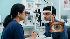
Diagnosis starts with a comprehensive patient history and exam of the eyes. Slit lamp study may show a look which is that of a beaten metal or silver finish of the back of the cornea also iris atrophy, nodules, adhesions or pupil displacement.
Gonioscopy involves looking into the drainage angle and also identifying peripheral anterior synechiae, which are of a cellular nature which may cause blockage of fluid outflow. Also we use Specular microscopy for the purpose of studying the endothelial cell pattern and at the same time may note down the characteristic light dark reversal of abnormal cells.
Confocal microscopy at times gives us cellular level images which we see in cases of uncertain diagnosis. Should corneal swelling which distorts the view of the angle occur, ultrasound biomicroscopy may play a role in the assessment of what is going on within the structure. When we see that the patient has glaucoma we also do studies of the optic nerve, perform retinal nerve fiber layer optical coherence tomography, run visual field tests and repeat pressure measures. Corneal thickness is a factor we look at because it does play a role in the interpretation of the eye pressure readings.
These studies determine if the iridocorneal endothelial syndrome is stable, slow to progress or if it is already at play in the optic nerve. Specialized imaging is useful because this rare eye disease may at times be missed when early iris changes are subtle, also it helps to tell the difference between ICD and bilateral corneal disorders.
The ophthalmologist may compare what we see with Fuchs endothelial corneal dystrophy, posterior polymorphous corneal dystrophy, Axenfeld Rieger spectrum, uveitis, iris melanoma and other corneal disorders. Unlike Iridocorneal Endothelial Syndrome which is a bit different in that it is mostly unilateral, these other diseases tend to be bilateral, inherited, or present at birth.
Treatment for ICE Syndrome
At present there is no treatment which reliably targets the root abnormal cell process in all patients. Iridocorneal endothelial syndrome we manage based on its complications which include high eye pressure and loss of corneal clarity.
A patient which has mild symptoms, normal intraocular pressure and good vision may at first require close monitoring as opposed to immediate surgery. In some cases the ophthalmologist will prescribe pressure reducing eye drops. Drugs which decrease production of aqueous humor are also an option we may try as the usual drainage routes may be blocked.
Patients should not begin, stop or switch glaucoma medications without first speaking to their health care provider. As this rare eye disease may affect corneal clarity and fluid outflow, treatment options should be based on a full picture more so than a single pressure reading.
Glaucoma in ICE syndrome is also a challenging issue which we see to be a result of variable membrane and angle adhesions. When medical options fail to reduce pressure enough, we may turn to procedures like trabeculectomy which includes the use of antifibrotic agents or the use of a glaucoma drainage device.
The choice of procedure depends on angle anatomy, prior surgery, state of the cornea and the extent of optic nerve damage. Long term follow up is required which includes monitoring for scar formation or re-growth of membranes that may in time nullify the results of the surgery. For the issue of corneal oedema, we may at first use what is called a symptomatic approach but in the case of end stage endothelial failure we may have to do a corneal transplant. Endothelial keratoplasty procedures like DSAEK or DMEK will replace the affected internal layers of the cornea while leaving most of the patient’s cornea in place.
In cases of complex eyes which present with extensive adhesions or other structural changes the surgeon may recommend a different approach. Transplant results are not guaranteed in such cases and continuous pressure monitoring is an issue post op.
In comparison to other corneal diseases this condition’s ongoing angle changes play a role in postoperative and daily eye care.
Regular review is at the core of managing iridocorneal endothelial syndrome which can progress even in between periods of stable vision. Each patient’s course with ICE syndrome is unique. Follow up intervals depend on eye pressure, corneal swelling, optic nerve appearance, visual field results and past treatment.
Patients should follow the prescriber’s instructions exactly for all eye drops and bring to all visits any eye medicine they are using. Also report right away if they have more pain, redness, halos, headache or a sudden change in vision.
Rubbing eyes out, use of left over steroid drops or purchase of pressure reducing medicines without consultation can delay proper treatment or cause other issues. This is a rare eye disease which requires very close follow up even when it appears that your day to day vision is not affected.
There is at present no diet, vitamin or exercise which has been proven to reverse abnormal endothelial growth. What we do know is that healthy lifestyle choices play a role in overall eye health, but they are not a substitute for specialist care, pressure tests or prescribed treatment.
When Should You Consult an Eye Specialist?
If you notice persistent blur in one eye, repeated morning haze, halos, an irregular pupil, new iris pigmentation or unexplained eye pressure, set up a medical exam.
Seek immediate care for sudden onset of very severe eye pain, nausea, and also very red eyes or acute vision loss. While these may not be specific signs of iridocorneal endothelial syndrome, they do point to glaucoma, active inflammation or other corneal disorders which require prompt attention.
A thorough evaluation is especially important when there is a difference in appearance between the right and left eye. By determining which symptoms are from the cornea, drainage angle, iris or optic nerve we are able to put forward more precise treatment options.
Conclusion
Iridocorneal endothelial syndrome is a rare but serious eye condition which sees abnormal corneal endothelial cells affect the cornea, iris and drainage angle. Symptoms may present very gradually with morning blur, halos or a change in pupil size. Primary complications include corneal decompensation and secondary glaucoma.
Although at present we do not have a universal cure for this rare eye disease, timely diagnosis and individualized care may help control eye pressure, manage corneal swelling and preserve what is left of the vision. It is also very important that anyone who notices one sided visual impairment, iris changes or recurring eye pain to take in to see an ophthalmologist at once instead of putting off a full scale ophthalmic work up.
Frequently Asked Questions
1. Is Iridocorneal Endothelial Syndrome Hereditary?
Iridocorneal endothelial syndrome which is to say is an acquired and sporadic condition and not a typical inherited disease. Family history is not usually present which in turn helps to differentiate it from certain inherited corneal disorders although only an eye exam can establish the right diagnosis.
2. Can ICE syndrome Affect Both Eyes?
ICE syndrome primarily affects one eye. While bilateral involvement does report, it is rare. Despite that, doctors still play it safe and examine both eyes because there may be a coexisting condition in the fellow eye.
3. Can the Condition Cause Blindness?
Untreated intraocular pressure increase or late stage corneal failure can cause severe and at times permanent visual loss. As is the case with other sight threatening corneal disorders the stage at diagnosis plays a role in what treatments are available. Early diagnosis, pressure control and treatment of corneal edema can reduce risk, but results also depend on the degree of the disease and response to treatment.
4. Is This Rare Eye Disease Curable?
This rare eye disease at present does not see a cure for the abnormal endothelial process in all patients. But we do have medications and surgeries which improve glaucoma, bring back some corneal clarity in some cases and may help in preserving useful vision.
5. Does Every Patient Need Surgery?
No. Mild form of Iridocorneal Endothelial Syndrome with invariable pressure and good vision which may be put on watch. This rare eye disease is treated based on what complications present themselves which may not be what the name suggests.
Surgery is considered when glaucoma is not adequately controlled, corneal swelling significantly affects vision or structural changes create another clinical problem.
6. Are ICE syndrome and Fuchs Dystrophy the Same?
No. ICE syndrome usually presents as a unilateral issue and has to do with abnormal iris and drainage angle changes. Fuchs endothelial corneal dystrophy is a more bilateral issue and presents a different clinical picture. In both conditions corneal swelling may present and that is why specialist testing is important.