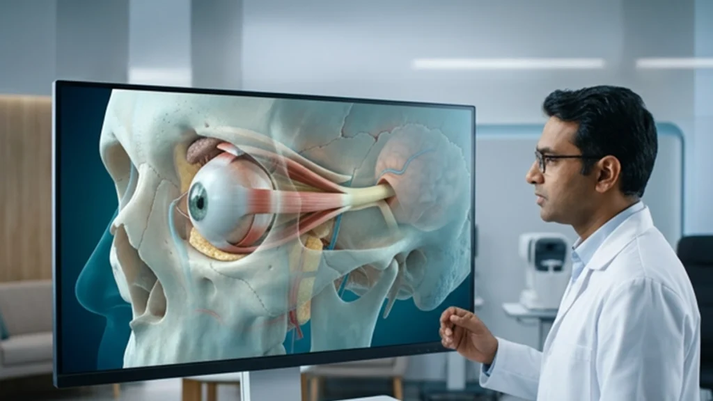
The human eye is a small organ but it is within a complex protective system. This system which we term the eye orbit supports the eyeball as well as related tissue which play roles in vision and eye movement. It includes bones, muscles, nerves, blood vessels, fat, and the lacrimal system. At the same time these components protect the eye and also allow for smooth movement.
We also refer to the eye orbit as the eye socket. But the socket is more than just a void in the skull. Studying its anatomy also helps to better understand injuries, infections, nerve problems and other orbital conditions.
What Is the Eye Orbit?
The eye orbit is a bony structure in the skull which includes the eye and its support systems. Each socket has a shape which tapers at the rear. The optic nerve which leaves the eye goes through a hole at the back of this main section. The socket contains the eye, extraocular muscles, nerves, blood vessels, tear duct structures, connective tissue, and orbital fat. The fat which is in there protects the eye, and the other structures which surround it keep the eye in place. This design gives the socket a protective and functional role.
Bones That Form the Eye Orbit
Seven which make up the eye orbit are the frontal, sphenoid, zygomatic, maxilla, palatine, lacrimal, and ethmoid bones. They in turn form the roof, floor, medial and lateral wall of the orbital socket.
At the same time that this bone structure of the eye socket is to be strong enough to protect the eye it also must provide for the passage of nerves and blood vessels.
| Orbital Area | Main Bones Involved | Main Role |
| Roof | Frontal and sphenoid | Separates the orbit from the cranial cavity |
| Floor | Maxilla, zygomatic and palatine | Supports the lower portion of the orbit |
| Medial wall | Ethmoid, lacrimal, maxilla and sphenoid | Forms the inner orbital boundary |
| Lateral wall | Zygomatic and sphenoid | Provides strong outer protection |
Damage to an eye socket bone can occur after falls, road accidents, sports injuries, or direct facial trauma. Some fractures can affect eye movement or alter the position of the eyeball.
Muscles Inside the Eye Orbit
The eye orbit contains six main extraocular muscles which control eye movement. They are the superior, inferior, medial and lateral rectus muscles and also the superior and inferior oblique muscles. Also present is the levator palpebrae superioris which raises the upper eyelid. These muscles allow for looking up, down, in, out, and diagonally. Also they are what allows both eyes to stay aligned. An issue with a muscle or its nerve supply may cause double vision or abnormal eye alignment.
Nerves of the Eye Orbit
In the orbital socket we find several key nerves. The optic nerve which is also known as cranial nerve II transmits visual info from the retina to the brain.
Cranial nerves III, IV, and VI which control various extraocular muscles. The oculomotor nerve which is also in that group of nerves controls the majority. The trochlear nerve supplies the superior oblique muscle and the abducens nerve which is also in this group of nerves controls the lateral rectus. Also we have the sensory branches of the trigeminal nerve serving the areas around the eye orbit.
| Nerve | Main Function in the Orbit |
| Optic nerve (CN II) | Carries visual signals to the brain |
| Oculomotor nerve (CN III) | Controls most eye movements and eyelid elevation |
| Trochlear nerve (CN IV) | Controls the superior oblique muscle |
| Trigeminal nerve (CN V1) | Provides sensation around the eye and forehead |
| Abducens nerve (CN VI) | Controls outward eye movement |
Because several nerves pass through a relatively compact eye orbit, inflammation, trauma, or compression may affect vision and eye movement.
Common Disorders Affecting the Eye Orbit
Orbital fractures can damage an eye socket bone after facial trauma. As for what area of the eye is affected and the degree of the injury a fracture may cause swelling, pain, double vision, limited eye movement, or a change in eye position. Orbital cellulitis is an infection of the eye’s tissue in orbit. It may cause painful swelling, redness, fever, reduced eye movement, or vision problems. Also because of the seriousness of an infection in the orbital socket prompt medical attention is important.
Thyroid eye disease also affects muscles and tissues in the eye orbit. There may be swelling which pushes the eyes out or which interferes with smooth eye movement. Also within the orbital cavity tumors and other abnormal growths may develop within the orbital socket put pressure on adjacent structures. We also see inflammation, vascular issues, nerve problems, and severe injuries may also affect the eye orbit. Symptoms will vary based on which tissue is affected.
When Should You See an Eye Specialist?
If you have developed sudden double vision, severe pain around the eye orbit, reduced vision, significant swelling, restricted eye movement, or a noticeable change in eye position go to see a doctor. After an injury which includes the eye socket bone see a doctor for an assessment. An ophthalmologist will test your vision, reaction of the pupils, movement of the eyes, and the nearby tissue. In the case of a deep orbital socket problem is suspected.
Conclusion
The eye orbit is a compact yet very organized part of the visual system. It’s bones which protect the eye, its muscles which cause movement, and its nerves which tie in vision and eye movement to the brain. Even a small issue in this area may affect nearby structures.
Persistent pain, swelling, double vision, restricted eye movement, or changes in vision should not be put off. An ophthalmologist is able to determine what in the eye socket is affected be it bone, nerve, muscle, or some other structure and will recommend the right treatment.
Frequently Asked Questions
- What is the eye orbit?
The eye orbit is a protective bony structure that includes the eye along with associated muscles, nerves, vessels, fat, and supporting tissues.
- How many bones form the eye orbit?
Seven bones make up the eye orbit frontal, sphenoid, zygomatic, maxilla, palatine, lacrimal, and ethmoid.
- Is the eye socket bone easy to fracture?
An eye socket bone can break with severe facial trauma. The base and sides of the orbit may also be affected by the direction and force of the impact.
- What is inside the orbital socket?
The orbital socket includes the eyeball, extraocular muscles, optic nerve, other cranial nerves, blood vessels, lacrimal structures, fat, and connective tissues.
- Can an orbital disorder affect eyesight?
Yes. A disorder within the eye orbit may affect the optic nerve, muscles, blood vessels, or the eyeball. Some conditions thus produce double vision, restricted movement, or reduced vision.