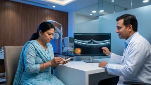
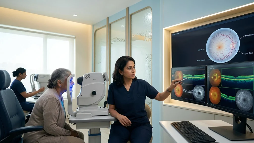
Retinopathy is damage to the retina, the light sensitive tissue at the back of the eye. It may result from diabetes, uncontrolled blood pressure, blood vessel disorders, certain medications or other health conditions. Of these Diabetic retinopathy requires special note as it may progress very silently. A retinopathy test is thus not something to put off until your vision has blurred it is a proactive assessment which may reveal early changes in small vessels before daily vision is affected.
For a person living with diabetes an early diabetic retinopathy diagnosis can put into play different treatment options which in turn may prevent avoidable sight loss. A full retinal examination is when the ophthalmologist looks at the macula, retinal blood vessels and optic nerve as opposed to just the power of the eye which is what we check for in a routine eye exam. That is important because by the time you notice retinopathy symptoms the structure has already changed. The National Eye Institute reports that in the early stages of diabetic retinopathy there may not be any symptoms and that the condition is diagnosed through a dilated eye exam.
What Does a Retinopathy Test Check?
The term retinopathy test is a broad one. It usually includes a series of tests which are chosen based on the patient’s health history, vision changes and suspected cause. The visit may start with visual acuity and pupil tests which are followed by drops to dilate the pupils. The doctor will then do a retinal examination which will look for things like bulged blood vessels, bleeding, fatty deposits, swelling, blocked circulation, abnormal new blood vessels or scar tissue.
In diabetes the goal is to achieve an accurate diabetic retinopathy diagnosis, determine if the macula is affected and to grade the severity. The Indian Ministry of Health and Family Welfare reports that in the early stage of diabetic retinopathy may go unnoticed and that screening is important to identify the disease before extensive visual damage. The fact that there are no retinopathy symptoms should not be taken as proof that the retina is healthy.
A routine vision test and a retinal exam are not the same. From a chart of letters we see how well a person is seeing at that time, but a retinopathy test looks at the tissues which receive light. It is possible for someone to do well on the chart but to have early vessel changes present. That is why a dilated retinal examination is key to screening and follow up, in particular for diabetics or people with long term high blood pressure.
Why Early Testing Matters
One of the key issues in diabetic retinal disease is that it develops very quietly. At first there may be retinopathy symptoms at all and minor changes which go undetected may not affect the person’s ability to read, drive or work. It is only when persistent blur, distortion or floaters present that the retina may be damaged. A person may present for care only once their vision has declined but by then leakage or bleeding may have already occurred. In the early stages a retinopathy test gives the ophthalmologist an opportunity to spot the damage and treat it before the patient’s vision breaks down.
A documented case of diabetic retinopathy diagnosis also serves as a base line. We will compare future photos and scans to the initial results to determine if the condition is stable or is progressing. This is a better approach than waiting for new retinopathy symptoms. Also follow up may bring to light the need for better coordination with the patient’s physician when blood glucose, blood pressure, cholesterol or kidney health is an issue.
The National Eye Institute reports that people with diabetes should have regular comprehensive dilated eye exams which typically are yearly although should be tailored to the person. During pregnancy or when there is macular swelling, advanced changes or previous treatment we may see more frequent evaluation. The interval between each retinopathy test is based on what is found at the visit and the ophthalmologist’s assessment.
Retinopathy Symptoms That Need Attention
Later retinopathy symptoms may present as blurred or variable vision, difficulty with small print, reduced color intensity, poor night vision, dark spots, floating forms or cobweb type marks. Some report straight lines as wavy or a void in the field of vision. While these symptoms do not in and of themselves confirm the disease, they are reason enough to schedule a retinopathy test.
A sudden onset of floaters, flashes, a curtain like shadow or acute vision loss requires prompt assessment. Such retinopathy symptoms may present with bleeding in the eye, traction on the retina or retinal detachment. The Ministry’s ophthalmology guidance reports progressive central vision loss, dark painless floaters and severe vitreous hemorrhage as common signs of advanced diabetic retinal disease.
Cataracts, glaucoma, retinal vein occlusion and age related macular disease present with similar symptoms. A structured retinal examination is used to tell these apart. A diabetic retinopathy diagnosis is based on what is seen in the retina at exam, imaging is used when needed to support the diagnosis which may not present with noticeable symptoms.
Tests Used to Examine the Retina
The doctor selects which retinopathy test for each patient based on the clinical question. Some patients require dilation and photography, while others will have high resolution scanning or dye based imaging.
| Test | What happens | What it helps assess |
| Visual-acuity assessment | The patient reads letters or symbols at set distances | Current clarity of vision and a baseline for comparison |
| Dilated fundus evaluation | Drops widen the pupils and the doctor views the back of the eye | A direct retinal examination for bleeding, deposits, vessel changes and new vessels |
| Fundus photography | A specialised camera records images of the retina | Documentation and comparison at later appointments |
| Optical coherence tomography | Light-based scanning creates cross-sectional images | Macular swelling and changes in retinal layers |
| Fluorescein angiography | Dye is injected and sequential photographs are taken | Leakage, blocked circulation and abnormal blood vessels |
| Tonometry | Eye pressure is measured | Associated pressure-related problems, including glaucoma |
Dilation is done to let in more light which in turn allows the doctor to see inside. Post procedure vision may be blurry and sensitive to light for a few hours which is why we recommend the use of sunglasses and we arrange for transportation home.
Fundus photographs produce a permanent record of the condition which we are looking at, also we have Optical Coherence Tomography or OCT that will tell us if there is fluid in or under the macula. Fluorescein angiography may be used in addition when the specialist wants to see if there is leakage, poor circulation or abnormal blood vessels.India’s guideline report includes photography in retinal screening and we use angiography for documentation of vessel changes and assessment of macular circulation. An in depth retinopathy test is useful if it answers a specific clinical question; it is not a routine requirement for all patients.
How Diabetic Retinopathy Is Diagnosed and Graded
The doctor will go over what type and which stage of diabetes the patient has, present medications the patient is on, past eye procedures, pregnancy status and other health issues. For the diabetic retinopathy diagnosis we look at the location and extent of retinal changes. What we term non-proliferative is when damage to blood vessels causes them to leak or close off without the growth of abnormal new ones. Proliferative disease is when these new fragile blood vessels do in fact grow which in turn increases the risk of bleeding and retinal detachment.
The macula is evaluated separately as swelling in this central area can cause loss of detail at various stages. A retinopathy test may report minor changes which are peripheral to the center but in the case of clinical macular edema those changes are of note. A careful retinal examination backed up by OCT or angiography when necessary helps the specialist to choose between watchful waiting and intervention.
| Finding or stage | What it may indicate | Usual management direction |
| No visible retinopathy | No diabetic vessel damage is seen at the visit | Continue diabetes care and attend the next advised retinopathy test |
| Mild to moderate non-proliferative changes | Small vessel bulges, tiny haemorrhages or deposits | Improve systemic control and monitor as advised |
| Severe non-proliferative disease | Widespread blockage and bleeding with greater progression risk | Closer follow-up and treatment planning |
| Diabetic macular oedema | Leakage causes swelling near central vision | OCT monitoring, injections or laser may be considered |
| Proliferative disease | New vessels raise the risk of bleeding and traction | Prompt injections, laser or surgery may be advised |
| Vitreous haemorrhage or tractional detachment | Blood or scar tissue affects vision and retinal position | Vitrectomy or another retinal procedure may be required |
A good diabetic retinopathy diagnosis will detail which eye is affected, whether the macula is involved, the stage of the disease and the schedule for the next retinal examination. Patients should request a simple explanation of the report and a set schedule for follow up.
Treatment Options After Diagnosis
Treatment is based on disease stage, macular involvement, present visual acuity and progression risk. At early diabetic retinopathy diagnosis it doesn’t always mean that ophthalmic intervention is required right away. In mild disease the ophthalmologist may recommend a watch and wait approach along with better control of blood glucose, blood pressure and cholesterol. The retinopathy test is then to be repeated at the recommended interval.
When macular oedema or abnormal vessels put vision at risk, medicines can be injected into the eye. Anti-VEGF treatment dampens the signals that contribute to leakage and new-vessel formation; for some patients, corticosteroids may be appropriate instead. Laser treatment can limit leakage or reach broader sections of oxygen-starved retina. The CDC identifies anti-VEGF injections as a first-line treatment for diabetic macular oedema, while the National Eye Institute includes injections, laser and surgery among established options.
Vitrectomy is a treatment we may turn to when bleeding doesn’t resolve on its own, or when scar tissue forms which in turn tugs at the retina, or when retinal detachment sets in. We can’t with this treatment reverse vision which has become permanently lost, instead we use it to prevent additional damage. By the time severe retinopathy symptoms can narrow the opportunity for timely care, and follow-up retinal examination remains necessary after injections, laser or surgery.
Preparing for the Appointment
Before a retinopathy test, bring in your current medication list, diabetes records and past eye reports. Also report to the clinic if you are pregnant, have kidney disease, are on blood thinners, have dye or drop allergies, also note down past reactions. Report when the visual changes started, if they are constant, and which eye or eyes are affected. This history is a support for diabetic retinopathy diagnosis but does not replace imaging and examination.
Reducing the Risk of Progression
No lifestyle change can do away with the need for routine retinopathy test but in terms of diabetic care which in turn affects retinal health keep your blood glucose at the target level set by your health care team, manage your blood pressure and cholesterol, stop smoking, take your medications as prescribed and keep up with your medical reviews. A normal retinal examination reports what we see at that time; it is not a guarantee that changes will not appear in the future. Also an improvement in retinopathy symptoms does not mean that leakage or swelling has resolved.
When to Seek Urgent Eye Care
Seek immediate care for sudden vision loss, many new floaters, flashes, a dark curtain in your field of vision, severe distortion or eye pain related to a vision change. A prompt retinopathy test and dilated exam will see if there is bleeding, retinal detachment, a vascular block or another emergency. A definite diagnosis must be made by an eye care professional; home vision charts and routine eye exams do not diagnose the retina.
Frequently Asked Questions
1. Is a retinopathy test painful?
A very common feature of retinopathy test is that they are very much tolerable. Dilation of the pupils with the use of drops may cause a stinging sensation which is short lived, also bright light used in the exam may be intense for a short while. For angiography and eye injections which are more in depth procedures the clinical team will go over what to expect before we do them.
2. Can diabetic retinopathy be present without symptoms?
Yes. Early in the course of the disease many times there are no retinopathy symptoms. Thus people with diabetes should present for their scheduled screenings instead of waiting for blurred vision which may not occur until the diabetic retinopathy diagnosis.
3. How often should the retina be checked?
Many folks that have diabetes are told to get a dilated retinal examination at least once a year but that may change according to what we see, pregnancy status, type of diabetes and treatment history.
4. Is retinal photography enough for every patient?
Photography is a useful tool for screening and documentation, but it may not provide all answers. Upon suspicion of swelling, reduced circulation or advanced disease the doctor may turn to OCT, angiography or another retinopathy test.
5. What happens after an abnormal result?
The results are by severity and macular involvement. We may do more close observation, better control of the system, injections, laser or surgery. The plan goes by the diabetic retinopathy diagnosis and the status of each eye.
6. Should I book an eye assessment when my blood sugar is controlled?
Yes. Good diabetes control reduces risk but does not do away with the need for a retinopathy test. Screening is to identify changes before vision is affected and to determine the appropriate monitoring interval.